February 20, 2018
SIR 2018 March 17 - 22 / Los Angeles, California - Los Angeles Convention Center
Read Here

30% of healthcare workers in U.S. hospitals are unvaccinated, large-scale analysis reveals
Nearly 30% of healthcare workers across the U.S. are still unvaccinated, according to a new
study spearheaded by the Centers for Disease Control and Prevention.
While
vaccination rates ballooned from 36% to 60% in the four-month period between January and
April of this year, they soon tapered off, reaching 70% by September.
The data, which
includes 3.3 million healthcare personnel (HCP) across more than 2,000 American hospitals,
was published Thursday in the American Journal of Infection Control. Those involved say it’s
the most comprehensive study of its kind to date.
PHOTO GALLERY: How COVID-19 Appears on Medical Imaging
The photo gallery in the link shows the variety of radiological presentations of COVID-19 (SARS-CoV-2) in medical imaging, including computed tomography (CT), radiograph X-rays, ultrasound, echocardiograms and magnetic resonance imaging (MRI). The radiology images show examples of typical COVID pneumonia in the lungs and the numerous complications the virus causes in the body in multiple organs, including the brain, kidneys, heart, abdomen and vascular system.
Read the Full Article*Proposal Calls for Educating, Empowering Radiologists to Address Healthcare Disparity
The advantages of digital X-ray systems have played a key part in their adoption; their speed and accuracy, as well as quick processing times, allow for significantly higher patient screening volumes than earlier. This has pushed companies to focus on product development and innovation. However, these systems are priced at a premium, which slows their greater adoption. Other factors such as declining reimbursements, lack of infrastructure, particularly in developing and underdeveloped countries, and potential risks associated with radiation exposure are also expected to hinder the growth of this market.
Read the Full Article*X-ray market is expected to reach USD 16.4 billion by 2026
The advantages of digital X-ray systems have played a key part in their adoption; their speed and accuracy, as well as quick processing times, allow for significantly higher patient screening volumes than earlier. This has pushed companies to focus on product development and innovation. However, these systems are priced at a premium, which slows their greater adoption. Other factors such as declining reimbursements, lack of infrastructure, particularly in developing and underdeveloped countries, and potential risks associated with radiation exposure are also expected to hinder the growth of this market.
Read the Full Article*Portable Digital Radiography: Tackling Today’s Sports Injury Epidemic
The COVID-19 lockdown wreaked havoc on professional athletes, and now we are witnessing the
fallout. But thankfully, when injuries do happen, professional teams have a way to quickly
diagnose, treat and manage them. It all starts with the right diagnostic
imaging.
There’s a new epidemic out there and it’s a pesky problem on every
professional athletic field, court and course: Sports injuries.
After months of
relative inactivity during the COVID-19 pandemic, professional athletes are finding their
bodies are being tested more than before. With gyms and even parks closed for everyone,
resuming competitive sports after a lockdown is taking an unexpected toll on pro
athletes—with sports injuries on the rise in virtually every league.
UAB utilizes new method of X-ray imaging that captures the body in motion
The University of Alabama at Birmingham’s Department of Radiology has added a new method of
X-ray imaging to the Kirklin Clinic of UAB Hospital that allows health care personnel to
observe the bones and organs while in motion.
This new technology is dynamic digital
radiography, also known as DDR, and it allows physicians to watch a patient’s motion of
lungs and diaphragm during breathing to better analyze and diagnose suspected lung nodules,
chronic obstructive pulmonary disease, known as COPD, and interstitial lung disease and to
determine whether the diaphragm is paralyzed. Doctors can also observe the full range of
joint motion to help evaluate suspected injuries.
Are radiologists regaining control of cardiac imaging?
The rate of coronary CT angiography (CCTA) exams performed by radiologists in U.S.
hospitals increased by
more than 300% between 2010 and 2019, indicating that radiologists could be regaining
control of cardiac
imaging, according to a September 30 study in Radiology: Cardiothoracic Imaging.
Why?
In part due
to reimbursement cuts and technological advances that shifted cardiac imaging procedures
from cardiology
offices to the hospital, wrote a team led by Dr. Russell Reeves of the Center for Research
on Utilization
of Imaging Service (CRUISE) at Thomas Jefferson University in
Philadelphia.
"Cardiologists are not
performing nearly as many myocardial perfusion scans as they were, and most are now being
done at hospital
outpatient imaging departments," Reeves said in a statement released by the RSNA.
ACR Association offers up $225,000 to help local radiologists
The American College of Radiology Association is now accepting applications for its new
$225,000 political fund, established to help local radiologists fight nonphysician
scope-of-practice gains.
ACRA first announced the effort back in July amid ongoing
concerns that provider societies representing nurse practitioners, physician assistants and
others are pursing the ability to operate independently.
“The ACRA is committed to
working with state radiological societies to proactively educate lawmakers and counter
future scope threats,” the college said in a Sept. 24 announcement.
Radiology department uncovers pain-inducing problems with at-home workstations
Scores of radiologists were forced to set up makeshift workstations last year as the
coronavirus pushed people into home offices. But many are not ergonomically correct, causing
more pain and potential injuries, new research suggests.
The University of
Pennsylvania’s Department of Radiology based the findings on a program piloting virtual
ergonomic consultations for rads employed before March 2020. About 60% of providers suffered
neck discomfort while 40% complained of lower and upper back pain.
Radiologists
spend most of their time sitting at a workstation, and one recent study shows nearly 90%
claim to have musculoskeletal symptoms. Short, 30-minute consultations can enhance off-site
work areas and decrease injury risks, UPenn authors wrote Sept. 27 in JACR.
6 issues radiology must address after COVID-19
The COVID-19 pandemic has proved challenging to both the academic and industry communities
in radiology, and there are at least six issues the two groups must address going forward,
according to a report published September 6 in Academic Radiology.
On May 7, the
Association of University Radiologists (AUR) held its annual academic-industry roundtable.
Sixteen radiologists from 12 academic departments and radiological societies and 14 industry
leaders from 11 companies participated. The panel identified the following six themes
radiology and industry must address post COVID-19.
Race is the most pronounced driver of delays in screening-detected breast cancer diagnosis
A New York-based startup aiming to root out misdiagnoses in radiology has raised $25 million
in new
funding, leaders announced Monday.
Covera Health said the Series C financing comes by
way of
private equity firm Insight Partners, with additional contributions from existing investors.
Founded in
2017, the analytics company has established a Radiology Centers of Excellence Program,
working with
payers, physicians and employers to steer patients to high-quality imaging. Covera made
headlines in 2019
when it partnered with Walmart to help the retail giant’s employees avoid “misguided and
unnecessary”
treatment stemming from imprecise image interpretation.
How can physician extenders benefit a radiology practice?
The term "physician extender" can include a radiologist assistant (RA), a physician
assistant (PA), or a nurse practitioner (NP). The rules for use of these nonphysician
providers (NPPs) are different for each one and they vary from state to state according to
their licensure laws.
In some cases, the practice may bill and be reimbursed
separately for the services of an NPP. Understanding the differences is key to getting
started with physician extenders.
A registered radiologist practitioner assistant,
abbreviated RA, RRA, or RPA, is trained initially as a radiologic technologist and then
achieves additional training and credentialing. The American Registry of Radiologic
Technologists (ARRT) certifies RRAs, who work under the supervision of a radiologist and
serve as an assistant to perform patient assessment, patient management, and selected
clinical imaging procedures.
Radiologists among the most in-demand health workers, earning No. 5 highest starting salary
Radiologists are among the most in-demand physician specialists in the U.S. and receive
some of the highest starting salaries, according to recent figures from Merritt
Hawkins.
A majority of requests performed by the healthcare recruiting firm were for
physician specialists as opposed to primary care providers, with radiology the third most
requested specialty behind only family medicine and nurse practitioners
(No.1).
Radiology was also linked to the second most search assignments and job
openings, labeled as “absolute demand” by Merritt Hawkins. This, in part, reflects the
rising need for rads and the growing use of imaging procedures.
“Demand for both
radiology and anesthesiology … are increasing, a clear sign that volume of medical
procedures is growing,” authors of the 38-page report wrote. “Whether it is a diagnosis or a
procedure, little happens in healthcare without an image.”
Radiologists express ‘serious concerns’ with new national practice standards superseding state laws
Radiologists and other physicians are expressing “serious concerns” with efforts to develop
national practice standards for docs and other providers that supersede state
scope-of-practice laws.
The nation’s largest healthcare delivery system recently
initiated this process, hoping to help expand access to care amid the COVID-19 pandemic. But
the American College of Radiology, American Society of Neuroradiology and others are
“dismayed” that the Department of Veterans Affairs has operated opaquely in running the
process.
In a Thursday letter to the VA’s leader, more than 100 physician groups
including the ACR asked the massive health system to tread carefully in potentially
expanding nonphysicians’ scope of practice.
Race is the most pronounced driver of delays in screening-detected breast cancer diagnosis
Race is the most pronounced driver of delayed breast cancer diagnosis during regular
screenings,
according to a new study in the Journal of the American College of
Radiology.
Mortality rates from
the disease have declined steadily over the last decade, thanks to national mammography
screening
programs, among other factors. But “substantial” barriers to screening and primary care
still exist, which
can result in delayed treatment and poor outcomes.
Analyzing data from more than 700
women
diagnosed with breast cancer in their Atlanta health system, Emory University researchers
found some stark
differences. Black women were twice as likely to experience total delays greater than 45
days compared
with white women. And those with such a long lag after screening were 1.6 times more likely
to die,
authors found.
New radiology virtual consult model shows promise
July 22, 2021 -- A group of U.S. researchers found that virtual consultations can improve
radiology's value by enabling face-to-face visits among radiologists, primary care
physicians (PCPs), and their patients, according to a study published July 20 in the Journal
of the American College of Radiology.
A group from Massachusetts General Hospital
(MGH) in Boston conducted a feasibility study of a new patient-centered care model based on
video consultations. The team found that almost all radiologists, PCPs, and most importantly
patients who participated in the virtual consultations were not only satisfied with the
visits but also indicated they would participate in them again.
"Virtual radiology
visits led to high patient and provider satisfaction and increased interest in the
availability of this model in the future," wrote the study authors, led by Dr. Dania Daye,
PhD, of MGH.
Orthopedic surgeons’ most common reasons for opting not to read radiology reports
There’s a common perception in imaging that orthopedic surgeons often skip reading
radiology reports and just go straight for the scans. Is this a stereotype, and if not, what
are the reasons keeping such subspecialists from digesting rads’
recommendations?
European imaging experts set out to explore these questions through
an anonymous survey of 81 orthopedists, detailed Monday in the European Journal of
Radiology. Their findings offer key clues to help improve musculoskeletal radiology services
and interdisciplinary collaboration.
“Most reasons for not consulting radiology
reports in this study were time related,” Ricardo Donners, with the Department of Radiology
at University Hospital Basel in Switzerland, and co-authors wrote July 19. “Accordingly, the
majority of recommendations for radiology service improvement addressed the time issue as
well, suggesting faster report turnaround times and shorter texts for improved reporting
practice.”
Hospital reduces radiology reporting disruptions, CT wait times with simple practice tweak
One radiology department is finding success reducing both interruptions during reporting
and patient wait times with a few minor practice tweaks, according to a study published
Monday.
Chelsea and Westminster Hospital particularly experienced starts and stops
during the vetting of plain computed tomography head scans and CT of the urinary tract. But
by taking these tasks off radiologists’ plates, the 430-bed London teaching hospital saw
incoming calls drop 30% for head scans while wait times fell 40%.
“Reducing
disruptions during radiology reporting has the potential to improve radiologists’ ability to
manage workload, job satisfaction, stress levels, efficiency, reporting accuracy and
therefore patient safety,” lead author Christopher Watura, a musculoskeletal radiology
fellow with the National Health Service at the time of the study, and colleagues wrote June
21 in Current Problems in Diagnostic Radiology. “Radiology has a pivotal role in most
in-patient care pathways and so smoothing scanning bottle necks is expected to contribute to
improved service effectiveness, including reduced patient length of stay.”
Perspectives on Interventional Radiology
A provider’s first-person encounter with the specialty as a patient.
I arrived
at the interventional radiology suite and was greeted by a man with a thick red beard. It
was beautiful. This patient had metastatic bone lesions to the spine causing him great pain.
Radiofrequency ablation was being used as a palliative measure.
I heard a lot about
interventional radiology and was very excited. I heard it was a very procedure-oriented
field and very innovation-driven. I knew everything about the patient. His history with
cancer was humbling. I wrote the various patient details on a piece of paper. I took one
last look at my paper, confidently donned gloves, and stepped up to the table. I was excited
to get a close-up view and engage for the next 5 hours.
However, the procedure
involved a few sticks and a few pokes. It was done in 30 minutes, and the patient was rolled
out to the post-procedure bay. In the post-procedure bay, I spent time with the patient. The
patient smiled, talked about his puppies, and talked about his wife. He was looking forward
to his upcoming Alaskan cruise with his grandchildren.
He was talking, lucid, and
healthy after having cancer cells melted out of his spine.
What has radiology learned from the COVID-19 pandemic?
The COVID-19 pandemic has had an unprecedented impact on radiology departments and private
practices around the world. Many imaging operations have experienced a sharp decline in
volume as patients have delayed elective imaging procedures.
As the pandemic
subsides, many radiologists are welcoming back patients, ramping up procedures, and gaining
back volumes to pre-COVID-19 levels. But patients want to feel safe and secure as they
return, so radiologists must implement practices and policies that ensure patient
safety.
Radiologists have to consider what the "new normal" will be going forward.
Odds are it will incorporate social distancing, patient screening, sanitation, novel forms
of communication, and changes in radiology workflow to create an environment that feels safe
to patients.
Model can predict risk for radiology appointment no-shows
Missed appointments can impact patient care and result in a significant revenue shortfall for
radiology departments. However, a machine-learning algorithm could help prevent some of
these no-shows, according to researchers from the University of Maryland.
After
training a machine-learning algorithm using data from over 4 million scheduled outpatient CT
and MRI appointments at their medical system, a team of researchers led by Dr. Steven
Rothenberg found that the algorithm yielded promising results when applied prospectively for
predicting a patient's risk for missing their radiology appointment.
COVID-19 pandemic forced big changes in radiology workflows
The COVID-19 pandemic forced major changes in radiology workflow, but the past year also had
some silver linings and lessons for the future, according to speakers in a May 24 session at
the Society for Imaging Informatics in Medicine (SIIM) annual meeting.
In a panel
discussion, radiology directors and vendor executives talked about the challenges their
respective workplaces endured throughout the pandemic, polling members in the SIIM audience
on what they also faced while looking toward the future.
"I'm so happy to see things
are returning back to normal," said panelist Sylvia Devlin, IT manager at the Radiology
Clinical Operations and eRadiology Center at Johns Hopkins Medicine. "I think 2021 and
beyond is going to be so much better."
American College of Radiology elects 1st Black president in its nearly 100-year history
Renowned imaging expert Beverly G. Coleman, MD, has been elected as president of the American
College of Radiology. She is the first African American to be named to this position in the
organization’s nearly 100-year history.
Coleman brings her experience as the first
director of fetal imaging at the Children’s Hospital of Philadelphia Center for Fetal
Diagnosis and Treatment. She sat on the ACR Board of Chancellors from 2014 to 2020 and is a
former president of the Society of Radiologists in Ultrasound.
To Embargo or Not to Embargo? No Consensus on Releasing Patient Radiology Reports
Institutions still do not have a standardized way to make radiology reports available to
patients, but providing earlier access could be beneficial for all parties
involved.
Despite a mandate to give patients timely and easy access to their
radiology reports, many healthcare institutions have a built-in delay or embargo for
releasing and making radiology reports available to patients. But, even that isn't a
standardized practice.
Under the 21st Century Cures Act, providers are required to
make electronic medical test reports, including those from radiologist, available to
patients once they’re finalized. But, few facilities are following this requirement to the
letter, according to a team of investigators from the Yale School of Medicine.
7 Tips for Funding Radiology
A radiologist and former Johns Hopkins president lays out guidance for how radiologists can
successfully raise money for department activities.
In a world where grant dollars
are increasingly hard to come by, more and more academic institutions – and their radiology
departments – are relying on the generosity of others to foot the bill for their
activities.
Chances are, you will be faced with a need to conduct fundraising at some
point during your career, and having at least a rudimentary understanding of how to do it
will be important.
William Brody, M.D., Ph.D., a radiologist who served as president
of Johns Hopkins University from 1996 to 2009 offered several tips for successful
fundraising in his recent April 28 letter in the Journal of the American College of
Radiology.
Radiologists miss 24% of interval breast cancers they could have caught on initial screening mammogram
Nearly a quarter of interval breast cancers could have been caught at the initial mammogram,
according to a new analysis published Monday in Academic Radiology.
Such cancer cases
typically crop up between two scheduled rounds of screening, for various reasons that could
include clinician oversight or aggressive growth. Wanting to better understand the factors
leading to these instances, overseas scientists recently dove into the data searching for
clues.
Altogether, researchers pinpointed 1,010 interval cancer cases recorded as
part of the BreastScreen Norway program between 2004 and 2016. About 24% (or 246) were
deemed missed at the initial screening, with an average time between exams of about 14
months. Possible remedies to address this issue could include shortening intervals or
implementing supplementary screening techniques, experts advised.
American College of Radiology updates imaging appropriateness criteria with 13 new topics
The American College of Radiology has added more than a dozen new topics to its imaging
appropriateness criteria while revising several more, officials announced on
Monday.
ACR’s update covers several clinical scenarios, such as breast imaging in
transgender patients, or staging and follow-up for primary vaginal cancer. The college
additionally revised five other topics, with all including a narrative, evidence table and
summary of relevant scientific literature.
“ACR Appropriateness Criteria creates
consistent behaviors for medical imaging and interventional radiology procedures for all
patients,” Mark Lockhart, MD, chair of the committee that oversees the criteria, said in a
statement. “By employing these guidelines, providers enhance quality of care and contribute
to the most efficacious use of radiology.”
“Tip of the Iceberg”: The Impact of COVID-19 Long-Haulers on Radiology
Patients who have long-lasting, lingering symptoms of COVID-19 could present a growing impact
on imaging services.
COVID-19 vaccine rates may be rising, but the pandemic hasn’t
gone anywhere. But, it isn’t just the virus that’s lingering – many previously infected
patients are still feeling the effects of the disease.
Diagnosing COVID-19-positive
patients during the early stages of the pandemic put a heavy weight on radiology, and,
according to Lillian Chiu, M.D., a radiologist with New York Medical College, if more
patients become “long-haulers,” patients who experience long-lasting symptoms, that pressure
on radiologists and imaging services could increase.
The WOEs of Radiology
When something you see on an image stops you in your tracks.
Chugging along through my
work list this past week, I was stopped in my tracks by a honking-big liver mass that
stymied my brain’s pattern-recognition circuits. I’ve come to think of such abnormalities as
WOE lesions (“What on Earth…?”).
A WOE differs from a “Whoa!” lesion, although this
particular mass qualified for the latter category, too: Big enough you could see it from
across the room, bad, and/or a striking change from prior comparison studies.
The
initial challenge with a WOE is knowing for sure that it isn’t something you’ve seen before.
Okay, you haven’t seen anything like it lately, but there are only so many abnormalities out
there, right? So the first thing that happens is a ransacking of the long-term memory for
something that might not even be in its cobwebby recesses.
Intelli-C manufacturer, NRT, nominated for Ernst & Young Entrepreneur of the Year
NRT has been selected as nominee in the Ernst & Young Entrepreneur of the Year
competition 2020. We are delighted with this appreciation, and as a group we reflect as
follows: “This recognition coming our way echoes precisely the spirit of the entire NRT
team, - always focused on innovation, continuous improvements in all of our processes, and
not least overcoming barriers that appear to block our progress.”
Find Out More
American College of Radiology joins fight against Humana’s much-maligned PET/CT payment restrictions
The American College of Radiology has joined the battle against a much-maligned decision
by
Humana to restrict coverage for PET/CT imaging.
Back in October, the nation’s
fourth
largest commercial insurer notified plan members that they would not be eligible for
positron emission tomography with concurrently acquired CT in many instances. Those
include
cardiac, gastric, esophageal or neurologic indications, along with total body PET/CT for
screening.
Humana claimed that the technology is “experimental” and ““not
identified
as widely used and generally accepted,” drawing swift rebuke from nuclear medicine
groups.
Now, the college is joining in and asking the Louisville, Kentucky-based payer to
reverse
course.
Johns Hopkins Medicine Expert Weighs Devastating Impact of COVID-19 on Healthcare Workers
The practice of interventional radiology is vulnerable to medical errors and malpractice
concerns and may require a renewed focus on patient safety, according a new analysis
published on Tuesday.
That’s partly because of the subspecialty’s broad scope and
rapidly evolving technology to support its physicians. Procedural complications account
for
about one-third of lawsuits against interventional radiologists, and worldwide data
suggest
that IR would benefit from rigorous process improvement, Boston University experts wrote
in
Radiology.
Read the Full
Article*
* will take you to an external website
Top Trend Takeaways in Radiology from RSNA 2020 - An Article by Melinda Taschetta-Millane and Dave Fornell
December 17, 2020 — The key trends observed at 2020 Radiological Society
of
North America (RSNA) meeting all
focused around COVID-19 (SARS-CoV-2) and the impact it has had on radiology. The
underlying
question throughout the conference was how can the industry take the information from
this
past year and learn from it?
Alan Pitt, M.D., a neuroradiologist at Barrow
Neurological Institute in Phoenix, Ariz., sees the pandemic as a catalyst for change,
and a
way to reset how radiologists view healthcare. “We are an enabler of value of public
health
— taking care of as many people as possible and giving the highest value possible,” he
stressed in a Philips Editor’s Roundtable during the conference. “It’s time to refocus:
How
many lives can we save? 2021 should be interesting. We need to keep up this level of
urgency.”
Read the Full
Article*
* will take you to an external website
Johns Hopkins Medicine Expert Weighs Devastating Impact of COVID-19 on Healthcare Workers
December 16, 2020 — During the COVID-19 pandemic, health care workers
have
been at the forefront of the battle against the life-threatening illness. Sadly, they
are
not immune to the effects of the disease. Many have contracted COVID-19, and some have
died.
In a paper published Dec. 4, 2020, in the journal PLOS One, Junaid Razzak,
M.B.B.S., Ph.D., director of the Johns Hopkins Center for Global Emergency Care, and his
colleagues estimated the impacts of COVID-19 on the U.S. health care community based on
observed numbers of healthcare worker infections during the early phase of the pandemic
in
Hubei, China, and Italy, areas that experienced peaks in COVID-19 cases before the
United
States.
Read the Full
Article*
* will take you to an external website
CMS Imaging welcomes Crystal Weiser
CMS Imaging welcomes Crystal Weiser to the Team!
Crystal will be our new Medical
Accounts Manager in Virginia. Crystal's first day will be Monday, December 14,
2020.
Please join us in welcoming Crystal!
Fujifilm Presents Enhanced DR Solutions with the Availability Glass-free DR detector with ISS Technology
November 20, 2020 — Fujifilm Medical Systems U.S.A., Inc., a leading provider of diagnostic imaging and medical informatics solutions, will demonstrate the FDR D-EVO III, an industry-first, glass-free digital radiography (DR) detector with Irradiated Side Sampling (ISS) technology, and the latest additions to Fujifilm's Clinica family digital x-ray suites - FDR Clinica X OTC and FDR Clinica FS – during the 2020 Radiological Society of North America (RSNA) annual meeting. Previewed for the first time at RSNA 2019, the FDR D-EVO III DR detector is now available in 14 x 17 and 17 x 17 sizes across the United States. Engineered to improve detective quantum efficiency (DQE) for clearer images at lower doses, the FDR D-EVO III is currently the lightest 14x17 detector on the market with patented ISS. The groundbreaking detector could be paired with Fujifilm's advanced image processing options, such as Virtual Grid and Dynamic Visualization II. "Fujifilm is continuing its commitment to deliver innovative solutions by working with customers to learn how we can provide technology solutions that address their needs," said Robert Fabrizio, director of strategic marketing, modality solutions, Fujifilm Medical Systems U.S.A., Inc. "The leap-forward design of FDR D-EVO III provides imaging departments with a smart solution to endure tough medical environments and minimize life cycle costs. Imaging departments will be able to invest in a smart solution, while providing leading-edge technology to their patients that is built to last well into the future." In addition to the FDR D-EVO III and Fujifilm's line of DR detectors and mobile DR solutions, Fujifilm will spotlight its newest digital X-ray suites during the virtual RSNA show, including:
Hopkins radiologists warn of unsuspected COVID-19 pneumonia during routine outpatient imaging
Johns Hopkins radiologists are warning about the possibility of unsuspected COVID-19
pneumonia cases cropping up during routine imaging exams, and the need for greater PPE
diligence in the coming months.
Experts from the Baltimore-based institution gave
the
examples of two recent encounters in which patients presented with no symptoms while
undergoing CT scans for other reasons. Both ended up having the novel coronavirus, and
Megan
Lee et al. believe the specialty must remain hyper-vigilant as it works through the
pandemic-induced backlog of imaging exams.
Tropical Storm Eta Advisory - Monday, November 9, 2020
CMS Imaging, Inc. is monitoring Tropical Storm Eta and will execute our Storm Contingency
Plan in Florida and Georgia, if necessary. Tropical Storm Eta has sustained 60 mph winds
at
less than 14 MPH approximately 210 miles from Cuba's western tip. Tropical Storm Eta
will
approach the Florida Gulf Coast later this week and possibly bring impacts from rain,
wind,
and storm surges.
Keeping our team members and their families safe during this
storm
is our primary concern. We stand prepared to maintain our high level of service to our
clients. Our Charleston Call Center is open Monday to Friday, from 8:30 am to 5 pm.
Customers seeking assistance outside regular business hours may obtain weekend service
by
calling our phone number, 800.867.1821, or via email at service@cmsimaging.com.
For those
who
require service after the storm, we will begin to dispatch our service engineers to
areas
affected by the storm once the federal, state, and local authorities have deemed it safe
to
return and travel in the areas affected by Tropical Storm Eta.
We urge all those
in
the expected path of Tropical Storm Eta to monitor the National Oceanic and Atmospheric
Administration (NOAA) at https://www.noaa.com.
For additional information on how
to
prepare for hurricanes, visit the Federal Emergency Management Agency's (FEMA) website
at https://www.Ready.gov/hurricanes.
Please contact us at info@cmsimaging.com with any questions
regarding
CMS
Imaging's Storm Contingency Plan.
CMS finalizes rule requiring insurers to disclose negotiated rates for radiology, other ‘shoppable’ services
The Centers for Medicare and Medicaid Services finalized a rule on Thursday, requiring
private payers to disclose negotiated rates for imaging and other services.
All
told,
the initial list covers 500 “shoppable services,” running from CT scans of the head and
brain, down to foot and ankle x-rays. The administration said its goal is to compel
consumers to seek out the best deals for their treatment.
RSNA garners more than 14,000 registrations
More than 14,000 people have signed up for the Radiological Society of North America’s
upcoming meeting, the group reported Monday. It’s the first online-only event in the
history
of the annual gathering.
Back in May, RSNA decided to cancel its in-person 106th
Scientific Assembly and Annual Meeting because of the COVID-19 crisis. It’s only done so
twice before, in 1943 and 1945, due to transportation and gasoline supply problems
surrounding World War II.
Fewer Breast Cancers Being Diagnosed During COVID-19 Pandemic
Although the importance of breast cancer screening should always be emphasized, the
current
issues surrounding the coronavirus disease 2019 (COVID-19) pandemic have led to a
decrease
in the number of women who are continuing to follow their usual mammogram
schedule.
According to experts, neglecting to have vital cancer screenings puts
an
individual at a greater risk. Moreover, recent research has shown that, at the onset of
the
COVID-19 pandemic, nearly 50 percent of the American women who are breast cancer
survivors
have experienced disruptions in their care, revealing critical flaws in disaster
preparedness within the nation’s current healthcare system.
Take Note: Even Emergency Rooms Are Turning to Telemedicine to Cope with the Pandemic
Adoption of telemedicine has been a controversial subject in urgent care. While some
operators have seen its benefits by way of increasing access for patients—and, over the
past
few months, in reducing risk of COVID-19 transmission—others have expressed concern that
patients may “self-diagnose” and demand prescriptions without the benefit of a full
examination.
Read the Full
Article*
* will take you to an external website
In-Home X-ray Improves Experience for Dementia Patients
Bringing mobile X-ray into nursing homes shortens the experience for patients living with
dementia,
helping them feel safe and calm.
Providers who have imaged patients with dementia
know that the
process can be delicate and complicated as many of these individuals can experience
fear,
anxiety, and
even anger when they find themselves in an unfamiliar environment. But, new research out
of
Denmark
indicates at-home X-ray can help solve this problem.
Investigators, led by J.M.
Jensen, from the
Health Sciences Research Center at the University College Lillebelt, launched an
initiative
in an nursing
home in 2018. Their goal was to determine whether patients with dementia could respond
better to X-ray
services provided in their normal living space.
Physicians are using medical imaging as a defense against malpractice claims, study finds
U.S physicians are, in fact, ordering medical imaging as a defense against malpractice
claims, according to a new analysis released on Wednesday.
Studying national
Medicare
imaging trends over a 12-year period and lining them up against lawsuit payouts, experts
from Emory University found a correlation. In particular, states with a high-risk level
of
litigation were positively associated with utilization of advanced imaging, researchers
wrote Aug. 19 in JACR.
“Although causality cannot be asserted from our
observational
study, positive associations between paid malpractice litigation and subsequent advanced
Medicare imaging utilization support the notion that U.S. physicians use medical imaging
as
a defensive medicine strategy,” concluded Alexander Villalobos, MD, with the Department
of
Radiology and Imaging Sciences at the Atlanta institution, and coauthors in health
policy
and economics. “Policymakers seeking to curb unnecessary healthcare spending should
carefully consider the direct and indirect impacts of medical malpractice litigation on
physician practice behaviors.”
Cancer vs. COVID: When a Pandemic Upended Cancer Care
Healthcare institutions, particularly hospitals, have long been seen as a tempting target
by cyber-criminals. Holding vast swathes of highly sensitive and valuable data, as well
as
having heavily interlinked IT systems and extensive use of IoT devices, modern
organizations
are both especially vulnerable and potentially highly lucrative should attacks be
successful. Indeed, unlike most industries, cyber-attacks have the potential to directly
endanger lives when it comes to healthcare.
Additionally, the scale and
pressurized
nature of the work in institutions like hospitals mean staff, focused on their critical
roles, are highly susceptible to making security errors that open the door to
cyber-criminals. Raj Samani, chief Scientist and fellow at McAfee, said: “Due to the
size
and nature of organizations within the healthcare industry, and the data they hold, our
health service is often a target for cyber-attackers. The scale and variety of attacks
are
continually growing and evolving, and the tactics cyber-criminals use can be a
combination
of traditional phishing and vulnerability exploitation.”
World Health Organization urges against using chest imaging to diagnose COVID-19
Add the World Health Organization to the growing list of experts advocating against the
use
of chest imaging to diagnose patients with COVID-19 in most cases.
The special
agency
of the United Nations just released its own set of recommendations on radiology’s role
in
assessing and managing those with the novel coronavirus. WHO does not endorse chest
imaging
for diagnosing the disease—unless lab tests are unavailable, or results are delayed—but
noted that scans can be a useful tool for managing COVID patients.
WHO said
several
countries have asked for guidance in the use of chest imaging during the pandemic, with
the
agency publishing the results of its literature review Thursday in Radiology. A recent
global survey conducted by the International Society of Radiology found “important
variations in imaging practices related to COVID-19” that needed to be addressed, the
authors noted.
Hurricane Isaias Advisory Friday, July 31, 2020
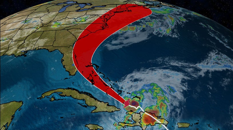
CMS Imaging, Inc. is monitoring Hurricane Isaias and will execute our Storm Contingency
Plan in Florida, Georgia, and the Carolinas if necessary. Hurricane Isaias is now a
Category
1 storm with sustained winds of 80 mph moving at less than 17 MPH approximately 15 miles
from the Great Inagua Island in the Bahamas. The forecast cone keeps the storm off the
east
coast of Florida and moving toward the Carolinas, but part of Central Florida is still
within the cone.
Keeping our team members and their families safe during this
storm
is our primary concern. CMS Imaging will continue to watch the situation very closely
and
will, if necessary, implement our Storm Contingency Plan. We stand prepared to maintain
our
high level of service to our clients. Our Charleston Call Center is open today, Friday,
July
31, 2020, and if the storm takes the projected path will be open as usual on Monday,
August
3, 2020. In the unlikely event, the storm takes a dangerous turn and threatens
Charleston;
we will close our call center and switch to a secondary location. In the event, our
secondary location is also affected; we have planned a third off-site call center
location
to continue to answer and respond to service calls. All call centers can be reached at
our
regular phone number, 800.867.1821, or via email at service@cmsimaging.com.
For those
who
require service after the storm, we will begin to dispatch our service engineers to
areas
affected by the storm once the federal, state, and local authorities have deemed it safe
to
return and travel in the areas affected by Hurricane Isaias.
We urge all those in
the
expected path of Hurricane Dorian to monitor the National Oceanic and Atmospheric
Administration (NOAA) at www.nhc.noaa.gov.
For additional information on how to
prepare
for hurricanes, visit the Federal Emergency Management Agency's (FEMA) website at https://www.Ready.gov/hurricanes.
Please contact us at info@cmsimaging.com with any questions
regarding
CMS
Imaging's Storm Contingency Plan.
American College of Radiology, RSNA ask feds to pump the brakes on autonomous AI
The two most prominent radiological societies in the U.S. are urging the federal
government
to proceed cautiously in its pursuit of artificial intelligence models that operate
autonomously.
RSNA and the American College of Radiology spelled out their
concerns
in a letter sent to the Food and Drug Administration on Tuesday. In it, top officials
from
the two groups said they believe it’s unlikely the FDA can provide assurances of such
technology’s safety in imaging care, absent further testing, surveillance and other
methods
of oversight.
They recommend that the FDA waits until such AI algorithms have a
broader penetration into the healthcare marketplace, prior to granting its approval in
the
future.
Effective Cybersecurity in Hospitals During #COVID19 and Beyond
Healthcare institutions, particularly hospitals, have long been seen as a tempting target
by cyber-criminals. Holding vast swathes of highly sensitive and valuable data, as well
as
having heavily interlinked IT systems and extensive use of IoT devices, modern
organizations
are both especially vulnerable and potentially highly lucrative should attacks be
successful. Indeed, unlike most industries, cyber-attacks have the potential to directly
endanger lives when it comes to healthcare.
Additionally, the scale and
pressurized
nature of the work in institutions like hospitals mean staff, focused on their critical
roles, are highly susceptible to making security errors that open the door to
cyber-criminals. Raj Samani, chief Scientist and fellow at McAfee, said: “Due to the
size
and nature of organizations within the healthcare industry, and the data they hold, our
health service is often a target for cyber-attackers. The scale and variety of attacks
are
continually growing and evolving, and the tactics cyber-criminals use can be a
combination
of traditional phishing and vulnerability exploitation.”
Model predicts 10,000 excess US deaths due to cancer screening delays: ‘We’re very worried’
AU.S physicians are, in fact, ordering medical imaging as a defense against malpractice
claims, according to a new analysis released on Wednesday.
Studying national
Medicare
imaging trends over a 12-year period and lining them up against lawsuit payouts, experts
from Emory University found a correlation. In particular, states with a high-risk level
of
litigation were positively associated with utilization of advanced imaging, researchers
wrote Aug. 19 in JACR.
“Although causality cannot be asserted from our
observational
study, positive associations between paid malpractice litigation and subsequent advanced
Medicare imaging utilization support the notion that U.S. physicians use medical imaging
as
a defensive medicine strategy,” concluded Alexander Villalobos, MD, with the Department
of
Radiology and Imaging Sciences at the Atlanta institution, and coauthors in health
policy
and economics. “Policymakers seeking to curb unnecessary healthcare spending should
carefully consider the direct and indirect impacts of medical malpractice litigation on
physician practice behaviors.”
9 Best Practices for Safely Re-Opening Mammography Services Post-COVID-19
After taking a nearly 70-percent hit to imaging volume during the COVID-19 pandemic,
mammography service lines are beginning to churn toward full volume again. It is a
process
that will look different for each practice or facility based upon their continued local
infection rates, as well as their size. But, according to industry leaders, there are a
set
of best practices that will help groups hit their post-COVID stride quicker.
To
this
point during the outbreak, the mammography landscape has looked very little like the
traditional environment with all screening services halted and many appointments
postponed.
And, there is scant indication that the return to “normalcy” will, indeed, be a re-start
of
business as usual.
How Harvard radiologists rapidly adopted a new reporting structure for possible COVID-19 patients
When a patient with possible COVID-19 presents at the radiology department, it can
trigger
a number of urgent steps that include isolation, testing, strict cleaning protocols and
the
use of scarce personal protective equipment. Wanting to provide physicians with a clear
process to report and launch these next steps, Harvard Medical School experts have
devised
an easy and “unambiguous” new reporting process for patients who possibly have the
disease.
Others—such as the Dutch Radiological Society—have previously designed
such
reporting and data systems. But those do not specifically address radiology department
workflows, including contact tracing and communication, experts wrote Thursday in JACR.
They’ve now implemented a “locally designed” reporting system at Brigham and Women’s
Hospital, with 71% of reports using the recommended terminology in the first two months.
RSNA Annual Meeting Goes Virtual for 2020
"As the convener of the world’s largest radiology meeting with over 50,000 attendees from 137 countries, our ability to conduct RSNA 2020 in Chicago is impacted by global public health considerations. With a mission that focuses on health and patient care, the primary consideration for RSNA is the health and safety of attendees, presenters, exhibitors, staff, and by extension, the global community. Therefore, we concluded it would be impossible to safely conduct RSNA 2020 in person and have decided to hold RSNA 2020: Human Insight/Visionary Medicine as an exclusively virtual event. The RSNA 2020 Virtual Meeting will be held completely online November 29 - December 5." - RSNA President James P. Borgstede, MD
Read the Full Statement*Opening Your Doors for Non-Emergent Care: Expert Guidance
For nearly two months, much of radiology’s imaging volume has been sitting on the
sidelines, waiting for the green-light for imaging centers to re-open their doors for
non-urgent scans. While there’s no exact date that will work for everyone to begin
elective
imaging again, the time is coming when facilities may feel comfortable doing
so.
But,
before you raise the curtain on the growing backlog of studies, there are many safety,
workflow, and personnel considerations you must address. To help you along the way, the
American College of Radiology (ACR) released guidance Wednesday, in the Journal of the
American College of Radiology, that can guide you to safely resuming non-emergent
imaging.
Coronavirus and the heart
Lung injury and acute respiratory distress syndrome have taken center stage as the most
dreaded
complications of COVID-19, the disease caused by the new coronavirus, SARS-CoV-2. But
heart
damage has
recently emerged as yet another grim outcome in the virus’s repertoire of possible
complications.
COVID-19 is a spectrum disease, spanning the gamut from barely
symptomatic infection
to critical illness. Reassuringly, for the large majority of individuals infected with
the
new
coronavirus, the ailment remains in the mild-to-moderate range.
Yet, a number of
those infected
develop heart-related problems either out of the blue or as a complication of
preexisting
cardiac disease.
A report from the early days of the epidemic described the extent of cardiac injury
among 41
patients
hospitalized with COVID-19 in Wuhan, China: Five, or 12 percent, had signs of
cardiovascular
damage. These
patients had both elevated levels of cardiac troponin — a protein released in the blood
by
the injured
heart muscle — and abnormalities on electrocardiograms and heart ultrasounds. Since
then,
other reports
have affirmed that cardiac injury can be part of coronavirus-induced harm. Moreover,
some
reports detail
clinical scenarios in which patients’ initial symptoms were cardiovascular rather than
respiratory in
nature.
Release of the “2019 Novel Coronavirus Detection Kit” Reduces labor and halves detection time
Shimadzu Corporation is announcing the release of its “2019 Novel Coronavirus Detection
Kit”, with sales
beginning on 20th April. For the time being the kits will only be available in Japan,
but
preparations are
underway to export these kits overseas from May onwards.
Current PCR methods for
the
detection of
the novel coronavirus (SARS-CoV-2) require the labor-intensive steps of RNA extraction
and
purification
from samples collected via nasopharyngeal swabs or similar. This presents an obstacle to
rapid detection
when dealing with large numbers of samples. This detection kit eliminates the steps of
RNA
extraction and
purification, significantly reducing the amount of work required to prepare samples, and
moreover halves
the overall time required for PCR detection from over 2 hours to approximately 1 hour.
When
using a PCR
device with the capacity for 96 samples, the workflow for all 96 samples can be
completed
within 1.5
hours. In addition, the omission of manual RNA extraction reduces the risk of human
error.
The
“2019 Novel Coronavirus Detection Kit” uses Shimadzu’s unique Ampdirect technology*1 and
has
been
developed based on the pathogen detection manual from the Japanese National Institute
for
Infectious
Diseases*2. This technology works to prevent proteins, polysaccharides, etc. contained
in
the biological
sample from inhibiting PCR, allowing the PCR reaction solution to be added directly to
the
biological
sample without the need for extracting and purifying DNA or RNA. Until now, Shimadzu has
developed and
released pathogen detection kits using Ampdirect technology for detection of
enterohemorrhagic E. coli,
salmonella, norovirus, etc., and is now applying this well-cultivated technology to
detection reagents for
the novel coronavirus.
*1: Ampdirect is a trademark of Shimadzu
Corporation.
*2: Japanese
National Institute for Infectious Diseases, “Pathogen Detection Manual for
2019-nCoV”
INTERPOL: #COVID19-Fighting Hospitals Facing Ransomware Deluge pandemic declaration
INTERPOL has been forced to issue an alert to global police about the heightened risk of
ransomware attacks on hospitals
and other front-line organizations as they battle the COVID-19 pandemic.
The
law
enforcement organization
said it issued a Purple Notice to all 194 member countries, highlighting the scale of
the
threat. Its Cybercrime Threat
Response team claimed to have detected a “significant increase” in attempted ransomware
attacks.
“As
hospitals and medical organizations around the world are working non-stop to preserve
the
well-being of individuals
stricken with the coronavirus, they have become targets for ruthless cyber-criminals who
are
looking to make a profit at
the expense of sick patients,” said Interpol secretary general Jürgen Stock.
CT should not be used as first-line tool against coronavirus, ACR warns following pandemic declaration
The American College of Radiology is urging physicians to only deploy computed tomography
in
very specific circumstances
to assess coronavirus patients. And CT “should not be used” to screen for—or as a
first-line
test to diagnose—the
virus.
ACR’s assertion comes the same day that the World Health Organization
declared the COVID-19 outbreak a
global pandemic. As of early Wednesday afternoon, there have been almost 122,000 cases
of
the disease, with 4,373
confirmed deaths. Another 66,000-plus individuals have also recovered from
it.
With limited availability of
viral testing kits and concerns about their specificity, some hospitals have taken to
using
chest x-ray or CT to assess
patients with the virus. However, ACR urged radiologists to use extreme caution in this
practice.
Read the Full
Article*
* will take you to an external website
CT should not be used as first-line tool against coronavirus, ACR warns following
pandemic
declaration
"CT Should Not Be Used As First-Line Tool Against Coronavirus, ACR
Warns
Following Pandemic Declaration". 2020.
Radiology Business.
https://www.radiologybusiness.com/topics/care-delivery/ct-scan-coronavirus-chest-x-ray-radiology-covid-19?utm_source=newsletter&utm_medium=RB-COVID19.
Is Anyone Paying Attention to Healthcare Security?
According to a recent report in TechCrunch, over one billion medical images from
patients
around the world — including
CT scans, X-Rays, ultrasounds — are available online for download to anyone with "an
internet connection and
free-to-download software.” It’s a pretty jarring number and while it is sure to get
people
talking, the question is:
will this revelation change anything?
More vulnerabilities are being found
in in
the healthcare space, and
yet very little action seems to come as a result. It’s a damning indictment on the state
of
digital risk management in
healthcare today, but the fact is that it’s not even surprising anymore.
While
more than 50% of healthcare
leaders report that “contending with fast evolving cyber threats” is the single greatest
challenge facing the industry,
32% still admit to never auditing their medical devices for known vulnerabilities!
Read the Full
Article*
* will take you to an external website
Source:
Oranski, Safi. 2020. "Is Anyone Paying Attention To Healthcare Security?".
Infosecurity Magazine.
https://www.infosecurity-magazine.com/opinions/paying-attention-healthcare.
Walmart Health opens second facility in Calhoun, Georgia
As reported in our"January 2020
Blog - Walmart Health enters the healthcare marketplace", Walmart cut
the
ribbon
on it's newest full-care clinic in Calhoun, Georgia with the asssitance of actor, Mark
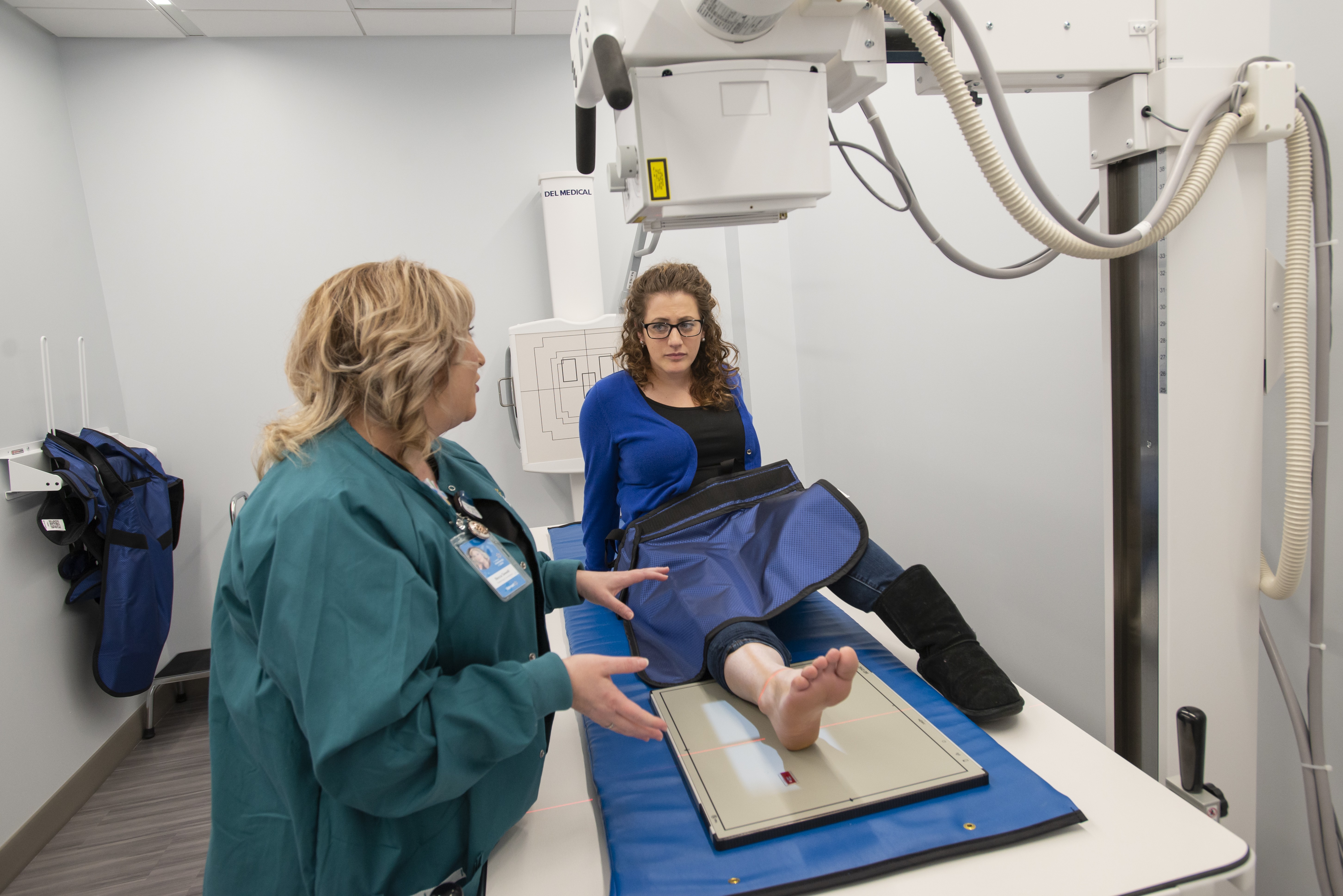
Wahlberg. The new Calhoun facility will offer primary and urgent care, labs, x-rays and
dignostics, counseling, dental,
optical, and hearing services as well as community health classes through a partenrship
with
Tivity Health. These
services are in addition to the full-service pharmacy already in the store. This new
urgent
care center will offer
upfront pricing for their services similiar to the previously-opened Walmart Health in
Dallas, Georgia. The upfront
pricing model will aloow patients to know exactly what the cost of services will be
prior to
making their appointment.
 In a time where many rural healthcare
facilities are on the brink of bankruptcies, Walmart is using their existing
facilities to ensure that the people of these communities have a easy access to
healthcare.

Agfa-Gevaert Group and Dedalus sign share purchase agreement for sale of part of Agfa HealthCare’s IT business
As announced on December 2, 2019, the Board of Directors of the
Agfa-Gevaert Group decided to
enter into exclusive negotiations with Dedalus Holding S.p.A. to
sell a part of Agfa
HealthCare’s IT business (the “Business”). Today, both parties
announce that they have
signed the share purchase agreement, under which Dedalus S.p.A.
would acquire 100% of the
Business at an enterprise value of 975 million Euro, subject to
regular working capital and
net debt adjustments.
The transaction is subject to
customary employees’
consultations, regulatory approvals and closing conditions. Both
parties aim to close the
transaction in the course of Q2 2020.
The Business
consists of the Healthcare
Information Solutions and Integrated Care activities, as well as
the Imaging IT activities
to the extent that these activities are tightly integrated into
the Healthcare Information
Solutions activities. This is the case mainly in the DACH
region, France and
Brazil.
(End of message)
About
Agfa
The Agfa-Gevaert Group develops, manufactures and distributes an
extensive range of analogue
and digital imaging systems and IT solutions, mainly for the
printing industry and the
healthcare sector, as well as for specific industrial
applications.
Agfa’s
headquarters and parent company are located in Mortsel,
Belgium.
The Agfa-Gevaert
Group achieved a turnover of 2,247 million euro in 2018.
Hospitals increasingly abandoning shielding in radiology as movement takes hold across US
On January 14th, 2020 Microsoft released patches for a
vulnerability that if exploited would
allow malicious software to appear as though it was from a
trusted, legitimate source. This
vulnerability bypasses Windows’ ability to verify cryptographic
trust, permitting an
attacker to create a false certificate to sign malicious code.
The NSA, in their
cybersecurity advisory, “assesses the vulnerability to be
severe” and the “consequences of
not patching the vulnerability are severe and widespread”. This
vulnerability could
facilitate easier spread of vulnerabilities like ransomware
undetected. Microsoft patches
ensure the cryptographic certificate is completely validated and
currently are the only
mitigation.
According to Microsoft, this vulnerability is
not being actively
exploited, although they assess this vulnerability likely to be
exploited in affected
operating systems. Patches have been issued for Windows 10,
Server 2016, Server 2019. A
Security bulletin will be issued shortly to provide guidance to
our field support channel
mitigate this vulnerability. This announcement will be updated
as necessary to advise of
further developments.
Shimadzu Medical Systems continually monitors for major
vulnerabilities such as these. We
will continue to monitor this threat and update this
announcement with any additional
product protection guidance or any known threat or exploit
information related to this
vulnerability. Additional information from Microsoft and the
security community are listed
below.
Hospitals increasingly abandoning shielding in radiology as movement takes hold across US
One prominent Chicago pediatric hospital is launching an “Abandon
the Shield” campaign to
begin moving away from using lead protection for patients during
imaging exams this spring.
Another a few miles away is mulling the same, despite the
initial “shock” clinicians felt
when presented with the idea.
Across the country, this
movement away from shielding
is beginning to take hold, Kaiser Health News detailed in a
prominent report issued Jan. 15,
and also published in the New York Times. At least a dozen U.S.
hospitals have already
changed their policies on protection during x-rays and many more
are starting to have the
conversation, one expert told the news outlet.
Doing so,
however, has not always
proven easy, with most healthcare consumers convinced that
shielding is necessary during any
imaging test.
Source:
Hospitals increasingly abandoning shielding in radiology as
movement takes hold across US
"Hospitals Increasingly Abandoning Shielding In Radiology As
Movement Takes Hold Across
US". 2020. Radiology Business.
https://www.radiologybusiness.com/topics/care-delivery/hospitals-abandon-shielding-radiology?utm_source=newsletter&utm_medium=rb_news
Hospital groups file lawsuit to block Trump's price transparency rule
U.S. hospital groups have challenged the Trump administration’s
rule that requires them to be
more transparent about prices they charge patients for
healthcare services, according to a
lawsuit filed on Wednesday.
The plaintiffs, including the
nonprofit American Hospital
Association (AHA), are looking to block the rule issued last
month that mandates hospitals
to publish pricing information of their services on the
internet.
“The rule ... does
not provide the information patients need. Mandating the public
disclosure of negotiated
charges would create confusion about patients’ out-of-pocket
costs, not prevent it,” the
plaintiffs said.
Source:
Hospital groups file lawsuit to block Trump's price
transparency rule
"Hospital Groups File Lawsuit To Block Trump's Price
Transparency Rule". 2020. U.S.. https://www.reuters.com/article/us-us-hospitals-idUSKBN1Y81YY.
Google AI system can surpass human experts in spotting breast cancer, study finds
Google’s artificial intelligence system can identify breast
cancer more accurately than
radiologists, according to a study published in Nature on
Wednesday.
The model,
created in collaboration with cancer researchers and Google
Health, was trained on X-ray
images, known as digital mammography, from women in the U.K. and
U.S. to spot signs of
breast cancer in the scans. Researchers used mammograms from
76,000 women in the U.K. and
more than 15,000 women in the U.S., according to Google Health.
Source:
Goodwin, Jazmin
2020. Usatoday.Com. https://www.usatoday.com/story/tech/2020/01/03/google-ai-system-can-beat-human-experts-spotting-breast-cancer/2795100001/.
Fujifilm to buy Hitachi's medical equipment business for $1.7 billion
According to a spokesperson for Fujifilm to Reuters, the global
imaging company is purchasing
Hitachi's diagnostic imaging line of service.
Fujifilm is
reportedly offering $1.7
billion for Hitachi's CT, MR, and Ultrasound line of products
and services. This purchase,
when finalized, will allow Fujifilm to challenge Siemens, GE,
Philips, and
Canon.
Check back for any additional details on this
industry-changing deal.
3 reasons why radiology leaders need to be on Twitter or risk falling behind
Radiology program directors must build an active presence on
Twitter or risk putting their
institutions at a “distinct disadvantage.”
,br>Imaging
experts from several institutions
across the country made that call to action in a recently
published opinion piece. They note
that numerous important functions of this job now often occur on
social media, from
networking to education, presenting opportunity for career
growth without straining their
budgets.
Surprise medical billing proposals could have ‘devastating unintended consequences’ on small radiology practices
Hundreds of small and independent physician practices are
concerned that congressional
proposals to address surprise medical bills could have other
“devastating unintended
consequences” on radiology and other specialties.
Almost
900 docs spelled out those
qualms in a sharply worded letter to U.S. House and Senate
leaders sent Thursday, Dec. 5.
Adopting too rigid of guidelines for payment could
unintentionally arm insurers that already
are using a “take-it-or-leave-it” approach to negotiations with
independent practices, they
wrote.
Canon Inc. Realigns Global Medical Business Strategy
TUSTIN, CA, December 2, 2019 – As part of Canon
Inc.’s global medical
business strategic realignment, the X-ray business operations
within Canon U.S.A., Inc.’s
wholly owned subsidiary Virtual Imaging, Inc. will transfer to
Canon Medical Systems USA,
Inc. (CMSU) effective January 1, 2020.
The global medical
imaging market is
anticipated to expand extensively year over year by 2025, fueled
by the aging population
coupled with the need for early stage detection of chronic
disease. As the leading medical
imaging market globally, the U.S. represents the largest
opportunity for future
growth.
Canon U.S.A., Inc., introduced the flat panel
detector into the U.S. market
in 1997 and Virtual Imaging, Inc. incorporated these detectors
into their portfolio of X-ray
systems. Today’s announcement of business realignment will build
on this strong legacy. This
transfer will help Canon Medical Systems USA, Inc., accelerate
additional opportunities for
growth while continuing to elevate the customer experience by
providing a single entity from
which to access significant technology advancements and purchase
equipment, as well as
obtain service and applications support.
As a result,
Canon Medical Systems USA,
Inc., will add vital digital radiographic and mobile offerings
to its current X-ray
portfolio of multi-purpose and fluoroscopic systems,
significantly expanding its product
line to be one of the broadest in the industry.
“This
strategic move offers customers
Canon Medical Systems USA, Inc.’s world-class and unified
experience for equipment
purchases, service and education solutions,” says Toshio
Takiguchi, senior managing
executive officer, Canon Inc., and president and CEO, Canon
Medical Systems Corporation. “At
Canon Medical Systems USA, Inc., our priority is always focused
on supporting our customers
by providing them with consistent, industry-leading X-ray
systems sales, service and
applications support.”
Learn more about Canon Medical’s
X-ray technology at this
year’s RSNA annual meeting in Chicago, December 1 – 5, 2019
(Booth #1933, South
Level).
About Canon Medical Systems USA, Inc.
Canon
Medical Systems USA, Inc.,
headquartered in Tustin, Calif., markets, sells, distributes and
services radiology and
cardiovascular systems, including CT, MR, ultrasound, X-ray and
interventional X-ray
equipment. For more information, visit Canon Medical Systems’
website at
https://us.medical.canon.
About Canon Medical Systems
Corporation
Canon Medical
offers a full range of diagnostic medical imaging solutions
including CT, X-Ray, Ultrasound,
Vascular and MR, as well as a full suite of Healthcare IT
solutions, across the globe. In
line with our continued Made for Life philosophy, patients are
at the heart of everything we
do. Our mission is to provide medical professionals with
solutions that support their
efforts in contributing to the health and wellbeing of patients
worldwide. Our goal is to
deliver optimum health opportunities for patients through
uncompromised performance, comfort
and safety features.
At Canon Medical, we work hand in
hand with our partners - our
medical, academic and research community. We build relationships
based on transparency,
trust and respect. Together as one, we strive to create
industry-leading solutions that
deliver an enriched quality of life. For more information, visit
the Canon Medical website:
https://global.medical.canon.
About Canon U.S.A.,
Inc.
Canon U.S.A., Inc., is a
leading provider of consumer, business-to-business, and
industrial digital imaging solutions
to the United States and to Latin America and the Caribbean
markets. With approximately $36
billion in global revenue, its parent company, Canon Inc.
(NYSE:CAJ), ranks third overall in
U.S. patents granted in 2018† and was named one of Fortune
Magazine's World's Most Admired
Companies in 2019. Canon U.S.A. is dedicated to its Kyosei
philosophy of social and
environmental responsibility. To keep apprised of the latest
news from Canon U.S.A., sign up
for the Company's RSS news feed by visiting
www.usa.canon.com/rss and follow us on Twitter
@CanonUSA.
Introducing new CXDI-702C and CXDI-402C Wireless Detectors from Canon Inc.
MELVILLE, NY, November 25, 2019 – Canon U.S.A.,
Inc., a leader in digital imaging solutions, is proud to
introduce the new CXDI-702C and CXDI-402C Wireless Detectors
from Canon Inc. These new detectors offer customers the superb
quality and reliability, as well as the features and
accessories, of the CXDI-710C, CXDI-810C and CXDI-410C Wireless
Detectors. Both the CXDI-702C and CXDI-402C Wireless Detectors
have received FDA 510(k) clearance for sale in the United States
of America1.
As seen by their open, curved
external cover, the CXDI-702C and CXDI-402C Wireless Detectors
were designed by Canon Inc. with usability in mind, helping to
facilitate both the user and patient experience. With the
lightweight handgrips, users are able to comfortably and easily
hold and handle the detectors on all four sides. Additionally,
the detectors were designed by Canon Inc. to provide certain
limited protection from dust and splashed water with limited
ingress of both permitted.
“We are excited to add the
CXDI-702C and CXDI-402C Wireless Detectors to the Canon U.S.A.
product portfolio as they align with Canon’s commitment to
developing high-quality diagnostic imaging devices with the goal
of helping to improve the quality of life for patients,” says
Tsuneo Imai, vice president and general manager, Healthcare
Solutions Division, Business Information Communications Group,
Canon U.S.A., Inc. “These detectors were designed by Canon Inc.
to meet clinical professionals’ demand for a cost-effective
detector technology with the ability to benefit multiple types
of diagnostic imaging facilities, and Canon U.S.A. is happy to
make them available to its customers.”
Canon U.S.A.
invites guests at Radiological Social of North America (RSNA)
Annual Meeting 2019, taking place Saturday, November 30, 2019
through Friday, December 5, 2019 at McCormick Place in Chicago,
to see the new CXDI-702C and CXDI-402C Wireless Detectors
firsthand in the Canon U.S.A. booth #3706. The booth will also
feature additional products from the Canon line of diagnostic
imaging devices.
For more information about Canon
radiography solutions available from Canon U.S.A., Inc., please
visit https://www.usa.canon.com/dr.
About Canon
U.S.A., Inc.
Canon U.S.A., Inc., is a leading
provider of consumer, business-to-business, and industrial
digital imaging solutions to the United States and to Latin
America and the Caribbean markets. With approximately $36
billion in global revenue, its parent company, Canon Inc.
(NYSE:CAJ), ranks third overall in U.S. patents granted in 2018†
and was named one of Fortune Magazine's World's Most Admired
Companies in 2019. Canon U.S.A. is dedicated to its Kyosei
philosophy of social and environmental responsibility. To keep
apprised of the latest news from Canon U.S.A., sign up for the
Company's RSS news feed by visiting www.usa.canon.com/rss and
follow us on Twitter @CanonUSA.
1
FDA clearance does not in any way denote FDA approval of
these devices.
Shimadzu Medical Systems releases the FLUOROspeed X1 edition RF system
Torrance, CA — November 18, 2019 ‐
Shimadzu Medical Systems USA, a
subsidiary of Shimadzu
Corporation, is proud to announce that they have released the
FLUOROspeed X1 edition,
patient side
conventional RF table system.
Shimadzu Medical Systems
USA
(SMS) has introduced a new
radiographic/fluoroscopic (RF) system called the FLUOROspeed X1
edition, a conventional RF
table system offering high image quality and a multitude
of features that improve work flow and operator efficiencies
thereby
contributing to lower
cost of care.
As the newest U.S. based product in the
FLUOROspeed series, the
FLUOROspeed X1 edition with its 665 lb. static patient weight
capacity (500 lb. all motion
weight capacity), easily performs
both bariatric and routine daily fluoroscopic and radiographic
exams. “The X1 is an
outstanding RF system
offering a costeffective balance of functionality to support a
wide
range of general RF
applications, such as chest,
abdomen, or extremities along with Upper GI’s, modified swallows
and
even joint injections”
says Charles Cassudakis, Director of Radiographic and RF
modalities
for SMS. “An
ambidextrous control
handle for the
imaging deck along with fingertip access to APR’s, image
recording
functions and
site‐specific
programmable function buttons, are all standard on the new X1 RF
system, all improving room
workflow,”
continues Cassudakis.
Jim Mekker, Product Director for
SMS
emphasizes that, “The X1
was designed and built from years of
collecting user feedback specifically from and for the US
marketplace and the result is a
remarkable, user
friendly and highly effective RF system with exclusive features
like
a park anywhere imaging
deck.
Additionally, the X1 is built with Shimadzu’s world renown
durability and reliability. This
system belongs
in every X‐Ray department nationwide.”
The FLUOROspeed X1 edition conventional RF system, designed with
patient side table controls
for the
operator, is practically priced and comes equipped with a
17”x17”
dynamic digital X‐ray
detector (FPD) in
the table bucky allowing it to both be used for fluoroscopy as
well
as radiographic exams.
With its 31.5‐
inch aperture opening between table top and deck, the X1 is the
ideal digital RF system
providing access
for imaging patients in wheelchairs, yet it can fit in smaller
rooms
where space is limited.
Furthermore, by
adding a second X‐ray tube on an overhead rail, the system
functionality and versatility of
the room
increases exponentially.
The FLUOROspeed X1 edition received FDA 510(k) clearance in
August
2019 and is now available
for sale
throughout the US.
About
Shimadzu
Shimadzu Corporation, founded
in
1875 in Kyoto, Japan and the parent of Shimadzu Medical Systems
USA
(SMS), is a global provider of medical diagnostic equipment
including conventional,
interventional and
digital X‐Ray systems. Shimadzu Medical Systems USA is
headquartered
in Torrance, California
with Sales
and Service offices throughout the United States, the Caribbean
and
Canada. Its sales and
marketing
office is located in Cleveland, Ohio, and its direct operations
has
headquarters in Dallas,
Texas. Visit
Shimadzu Medical Systems USA at www.shimadzu‐usa.com or call
(800)
228‐1429.
American Lung Association finds ‘dramatic’ increase in lung cancer survival rates, with radiologists playing a key role
Receiving that initial lung cancer diagnosis is not the death
sentence that it used to be for some Americans, with more
surviving the disease than ever before.
The American Lung
Association made that assertion in its newest State of Lung
Cancer report, released on Wednesday, Nov. 14. Almost 22% of
individuals diagnosed with lung cancer are still fighting five
years later, up from 17.2% just a decade ago. That’s a 26%
increase according to the ALA, one that’s partly attributable to
better screening using low-dose CT scans to catch the cancer
early on.
ACR ‘disappointed’ as feds finalize policy that could cost field billions
Radiology industry advocates expressed disappointment on Friday,
Nov. 1, after federal officials finalized a policy that industry
advocates said could cost the imaging field billions.
The
Centers for Medicare & Medicaid Services recently released the
final Medicare Physician Fee Schedule for 2020, which will take
effect on Jan. 1. One aspect of the now finalized rule has drawn
ire from the American College of Radiology, which estimated that
changes could cut radiology payment by $450 million in one year
alone, and $5.6 billion over the next decade.
Reject Analysis: Pictures or it Didn’t Happen
As a radiographer working in general radiology, you will be familiar with famous sentences like ”let me take one more, just to be sure” or “I don’t think this is lateral enough to visualize the pathology.” If you work in a radiology department, you’ll certainly have experienced some of these cases in your professional life:
ACR publishes updates to appropriateness criteria
The American College of Radiology (ACR) has added four new topics
and 15 revised topics to the ACR Appropriateness
Criteria.
The four new topics explore major blunt trauma,
pancreatic cysts, pneumonia in immunocompetent children and
suspected placenta accreta spectrum disorder. Revised topics
include dementia, female infertility, hemoptysis and more.
At RSNA 2019, Agfa Truly Transforms Digital Radiography, Through Its MUSICA Digital Nerve Center
MORTSEL, Belgium, Sept. 17, 2019 /PRNewswire/ -- All Agfa DR solutions delivered with the MUSICA Digital Nerve Center provide efficiency and intelligence – driven by artificial intelligence (AI) and deep learning*.
ACR ‘disappointed’ as feds finalize policy that could cost field billions
“Fast MRI” scans, which use quicker imaging techniques and don’t require sedation or
ionizing
radiation, can identify traumatic brain injuries (TBIs) in young patients, according to
a
new study published in Pediatrics.
The authors explored data from more than 200
patients recruited from June 2015 to June 2018. All patients were six years old or
younger
and also underwent head CT scans.
Read the Full
Article*
* will
take you to an external website
Hurricane Dorian Advisory 4 - September 6, 2019

CMS Imaging, Inc. continues to monitor Hurricane Dorian and is executing our Storm
Contingency Plan in North Carolina. Hurricane Dorian is now a Category 1 storm with
winds
exceeding 90 mph and is currently traveling along the Outer Banks of North Carolina. The
storm has created a large number of power outages in Florida, Georgia, and South
Carolina.
Our Charleston and Jacksonville Offices are open with limited staff.
Our
Charleston Call Center is currently taking calls at our regular phone number, 800.867.1821, or via email at service@cmsimaging.com.
For those who currently
require
service, we will assess the area, and if safe, we will dispatch our service engineers in
order of contact. For those areas still affected by the storm, we will begin dispatching
our
field service engineers once the federal, state, and local authorities have deemed it
safe.
Please contact us at info@cmsimaging.com with any questions regarding CMS
Imaging's
Storm Contingency Plan.
Hurricane Dorian Advisory 3 - September 4, 2019

CMS Imaging, Inc. continues to monitor Hurricane Dorian and is executing our Storm
Contingency Plan in Florida, Georgia, South Carolina, and North Carolina. Hurricane
Dorian
is now a Category 2 storm with winds exceeding 110 mph and is currently traveling
northwest
along the northeastern coast of Florida. The storm is expected to stay out to sea but
will
bring tropical-storm-force or hurricane-force winds to Florida, Georgia, South Carolina,
and
North Carolina.
To ensure the safety of our team members and their families, CMS
Imaging's Jacksonville Office is closed, and our Charleston Office and Call Center will
close today at 12:00 pm Eastern Daylight Time. We will transfer all of our calls to our
secondary off-site call center starting at noon. In the event, our secondary location is
also affected; we have planned a third off-site call center location to continue to
answer
and respond to service calls. All call centers can be reached at our regular phone
number,
800.867.1821, or via email at service@cmsimaging.com.
Based on the forecast from
the
National Weather Service, we expect to reopen our Jacksonville Office on Thursday and
our
Charleston Office and Call Center on Friday.
For those who require service after the
storm, we will begin to dispatch our service engineers to areas affected by the storm
once
the federal, state, and local authorities have deemed it safe to return and travel in
the
areas affected by Hurricane Dorian.
Please contact us at info@cmsimaging.com with
any
questions regarding CMS Imaging's Storm Contingency Plan.
Hurricane Dorian Advisory 1 - September 2, 2019
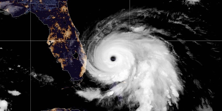
CMS Imaging, Inc. continues to monitor Hurricane Dorian and has begun executing our Storm
Contingency Plan in Florida, Georgia, South Carolina, and North Carolina. Hurricane
Dorian
is now a Category 3 storm with winds exceeding 120 mph and is currently stationary off
the
coast of the Bahamas before it's expected travel up the East Coast. The storm is
expected to
stay out to sea but will bring tropical-storm-force or hurricane-force winds to Florida,
Georgia, South Carolina, and North Carolina.
To ensure the safety of our team
members and their families, CMS Imaging's Charleston Office and Call Center will close
today
at 12:00 pm Eastern Daylight Time. We will transfer all of our calls to our secondary
off-site call center starting at noon. In the event, our secondary location is also
affected; we have planned a third off-site call center location to continue to answer
and
respond to service calls. All call centers can be reached at our regular phone number,
800.867.1821, or via email at service@cmsimaging.com.
For those
who
require service after the storm, we will begin to dispatch our service engineers to
areas
affected by the storm once the federal, state, and local authorities have deemed it safe
to
return and travel in the areas affected by Hurricane Dorian.
The CMS Imaging,
Inc.
Executive team will meet again tomorrow, and another Storm Alert will be issued.
Please contact us at info@cmsimaging.com with any questions regarding CMS
Imaging's
Storm Contingency Plan.
Hurricane Dorian Advisory 1 - September 2, 2019

CMS Imaging, Inc. is monitoring Hurricane Dorian and will execute our Storm Contingency
Plan
in Florida, Georgia, and the Carolinas if necessary. Hurricane Dorian is now a Category
5
storm with winds exceeding 180 mph moving at less than 5 MPH up the east coast of
Florida.
There is a level of uncertainty if the hurricane will make landfall, and if so, where it
will come ashore.
Keeping our team members and their families safe during this
storm
is our primary concern. The governors of Florida, Georgia, South Carolina, and North
Carolina have issued State of Emergency orders. Additionally, the governors of Georgia
and
South Carolina issuing Mandatory Evacuations along their respective coasts.
CMS
Imaging is watching the situation very closely and will if necessary, close our
Charleston
and Jacksonville offices if the storm presents a danger to our employees. Our Storm
Contingency Plan is in effect, and we stand prepared to continue our high level of
service
to our clients. Because of the Labor Day holiday, our Charleston Call Center is closed
today, Monday, September 2, 2019. In the event, the storm takes a dangerous turn and
threatens Charleston; we will close our call center and switch to a secondary location.
In
the event, our secondary location is also affected; we have planned a third off-site
call
center location to continue to answer and respond to service calls. All call centers can
be
reached at our regular phone number, 800.867.1821, or via email at
service@cmsimaging.com.
For those who require service after the storm, we will
begin
to dispatch our service engineers to areas affected by the storm once the federal,
state,
and local authorities have deemed it safe to return and travel in the areas affected by
Hurricane Dorian.
We urge all those in the expected path of Hurricane Dorian to
monitor the National Oceanic and Atmospheric Administration (NOAA) at https://www.noaa.com.
For additional information on
how
to prepare for hurricanes, visit the Federal Emergency Management Agency's (FEMA)
website at
https://www.Ready.gov/hurricanes.
The CMS Imaging,
Inc.
Executive team will meet again tomorrow, and another Storm Alert will be issued.
Please contact us at info@cmsimaging.com with any questions regarding CMS
Imaging's
Storm Contingency Plan.
Docs Brace for Medicare 'Appropriate' Imaging Rule
An article by Nicole Lou, Contributing Writer, MedPage Today
As the medical community braces for implementation of the Protecting Access to Medicare
Act
(PAMA) by the Jan. 1, 2020 deadline, some wonder if it's even feasible or if another
program
delay is on the horizon.
The policy, aimed at reducing unnecessary testing,
mandates
that all advanced diagnostic imaging orders go through an algorithm that provides key
confirmation codes required when Medicare is billed later on for the
service.
Dubbed
a "clinical decision support mechanism" (CDSM), this software processes each CT, MRI,
nuclear medicine, and PET order before spitting out its verdict to the ordering
professional: "appropriate," "maybe appropriate," or "rarely appropriate," according to
a
certain set of appropriate use criteria (AUC).
Read the Full
Article*
* will
take you to an external website
Mobile C-arm Technology Helps Build Efficiency
An article by Paramjit “Romi” Chopra, M.D.
A mobile C-arm is a medical imaging device that is based on X-ray technology and can be
used
flexibly in various ORs within a clinic. The name is derived from the C-shaped arm used
to
connect the X-ray source and X-ray detector to one another. Since the introduction of
the
first C-arm in 1955, the technology has advanced rapidly. Today, mobile imaging systems
are
an essential part of everyday hospital life; specialists in fields such as surgery,
orthopedics, traumatology, vascular surgery and cardiology use C-arms for intraoperative
imaging. The devices provide high-resolution X-ray images in real time, allowing the
physician to monitor progress at any point during the procedure and immediately make any
adjustments that may be required. Consequently, the treatment results are better and
patients recover more quickly. Hospitals benefit from cost savings through fewer
follow-up
operations and from minimized installation efforts.
Read the Full
Article*
* will
take you to an external website
Shimadzu Medical Systems receives FDA 510(k) for the FLUOROspeed X1 RF system
Torrance, CA — August 8, 2019 - Shimadzu Medical Systems USA, a
subsidiary
of Shimadzu Corporation,
is proud to announce that they have received FDA 510(k) clearance for the FLUOROspeed
X1,
patient
side conventional RF table system.
Shimadzu Medical Systems USA (SMS) has
introduced
a new radiographic/fluoroscopic (RF) system
called the FLUOROspeed X1, a conventional RF table system offering high image quality
and a
multitude
of features that improve work flow and operator efficiencies which contributes to lower
cost
of care.
As the newest U.S. based product in the FLUOROspeed series, the
FLUOROspeed
X1 with its 665 lb.
static patient weight and 500 lb. all motion weight, easily performs both bariatric and
routine daily
fluoroscopic and radiographic exams. “The X1 is an outstanding RF system offering a
cost‐effective
balance of functionality to support a wide range of general RF applications, such as
chest,
abdomen, or
extremities along with Upper GI’s, modified swallows and even joint injections” says
Charles
Cassudakis,
Director of Radiographic and RF modalities for SMS. “An ambidextrous control handle for
the
imaging
deck along with fingertip access to APR’s, image recording functions and site‐specific
programmable
function buttons, are all standard on the new X1 RF system, all improving room
workflow,”
continues
Cassudakis.
Jim Mekker, Product Director for SMS emphasizes that, “The X1 was
designed and built from years of
collecting user feedback specifically from and for the US marketplace and the result is
a
remarkable,
user friendly and highly effective RF system with exclusive features like a park
anywhere
imaging deck.
Additionally, the X1 is built with Shimadzu’s world renown durability and reliability.
This
system belongs
in every X‐Ray department nationwide.”
The FLUOROspeed X1 conventional RF system,
designed with patient side table controls for the
operator, is practically priced and comes equipped with a 17”x17” dynamic digital X‐ray
detector (FPD)
in the table bucky allowing it to both be used for fluoroscopy as well as radiographic
exams. With its
31.5‐inch aperture opening between table top and deck, the X1 is the ideal digital RF
system
providing
access for imaging patients in wheelchairs, yet it can fit in smaller rooms where space
is
limited.
Furthermore, by adding a second X‐ray tube on an overhead rail, the system functionality
and
versatility
of the room increases exponentially.
The FLUOROspeed X1 received FDA 510(k)
clearance
on August 5th, 2019 and is now available for sale
throughout the US.
About Shimadzu
Shimadzu Corporation,
founded
in 1875 in Kyoto, Japan and the parent of Shimadzu Medical Systems
USA (SMS), is a global provider of medical diagnostic equipment including conventional,
interventional
and digital X‐Ray systems. Shimadzu Medical Systems USA is headquartered in Torrance
California with
Sales and Service offices throughout the United States, the Caribbean and Canada. Its
sales
and
marketing office is located in Cleveland, Ohio, and its direct operations has
headquarters
in Dallas,
Texas. Visit Shimadzu Medical Systems USA at www.shimadzu‐usa.com or call (800)
228‐1429.
FDA Issues Draft Guidance on Medical Device Safety in MRI Environment
The U.S. Food and Drug Administration (FDA) issued a new draft guidance titled Testing
and
Labeling Medical Devices for Safety in the Magnetic Resonance (MR)
Environment.
The
MR environment presents unique safety hazards for patients and other persons with
medical
devices near or inside an MR system. This draft guidance, when finalized, is intended
to:
Is this imaging technique the future of prostate cancer treatment?
A newer imaging technique, PSMA PET/CT, may be able to identify the location of prostate
cancer recurrence better than traditional methods, according to new findings published
by
The Lancet Oncology.
PSMA PET/CT, or prostate-specific membrane antigen imaging,
is
not yet approved by the FDA, but researchers have been exploring its potential in recent
years. A team from the UCLA Jonsson Comprehensive Cancer Center in Los Angeles compared
the
performance of PSMA PET/CT with the treatment currently recommended for locating
prostate
cancer recurrence, 18F-fluciclovine PET/CT.
Read the Full
Article*
* will
take you to an external website
AHRA 2019: What it takes to become an ACR Diagnostic Imaging Center of Excellence
Earning the American College of Radiology (ACR) Diagnostic Imaging Center of Excellence
(DICOE) designation can help imaging providers stand out among the competition—if
they’re
willing to do what it takes to achieve that goal. A two-part presentation at the AHRA
2019
Annual Meeting in Denver, Colorado, examined the DICOE assessment process from the
perspective of a hopeful applicant and a medical physicist who helps perform on-site
evaluations.
Read the Full
Article*
* will
take you to an external website
CMS Imaging announces the first clinical installation in the US of the Intelli-C at ImageCare, LLC
North Charleston, SC, July 22, 2019 - CMS Imaging, Inc is proud to
announce
the first clinical installation in the United States of the Intelli-C at ImageCare, LLC.
The
Intelli-C, which is distributed nationally by Alpha Imaging, LLC, is a multipurpose
tilting
C fluoroscopic imaging system that offers true versatile imaging explicitly designed to
accommodate the needs of busy healthcare facilities, imaging centers, or hospitals. The
Intelli-C now offers ImageCare, LLC the extended ability to provide their patients
low-dose,
high-quality images for procedures ranging from radiographic, serial, and fluoroscopic
X-ray
procedures all on one system.
"We have been working with the Intelli-C for a few
months now. It provides all that I need in my practice of diagnostic and interventional
radiology. The configuration allows me to perform interventional procedures,
gastrointestinal procedures, pain management applications, and orthopedic interventions,
with low-dose radiation, without compromise in image quality. Even at low pulse rate
fluoroscopy, there is excellent visualization of target structures," said Dr. Timothy P.
Close, M.D. of Image Care, LLC. "If an imaging center or small hospital has only one
fluoroscopy or procedure room, they should have this equipment. In a busy hospital
setting,
this room provides flexibility and a broad spectrum of applications to complement
diagnostic
and therapeutic procedures," Dr. Close continued.
For more information on the
Intelli-C, visit – https://intelli-c.cmsimaging.com.
About
CMS
Imaging, Inc.
CMS Imaging, Inc. is the premier healthcare solutions provider specializing in the sales
and
service of diagnostic medical imaging equipment. Founded in 1987 in Charleston, SC as an
independent service organization, CMS Imaging has expanded its product line to include
MRI,
CT, Digital X-Ray, Advanced Fluoroscopic Systems, Software, and Informatics. For more
information, please contact CMS Imaging at 800.867.1821 or
info@cmsimaging.com.
About
ImageCare, LLC
ImageCare, LLC is Northeast Columbia's (South Carolina) only full-service outpatient
radiology facility. We specialize in diagnostic procedures and provide a wide range of
imaging services. Our radiologists are fellowship-trained and board-certified by the
American Board of Radiology. ImageCare provides a written report to the referring
physician's office within 24 hours and offers physician-to-physician call reports in
cases
that involve significant abnormalities.
About Alpha Imaging,
LLC
Alpha Imaging, LLC headquartered in Willoughby, Ohio, was established in 1986 and is one
of
the country's largest independent distributors of medical imaging equipment and related
products and services. For more information, please contact Alpha Imaging at 800.331.7327 or info@alpha-imaging.com.
MedCurrent Announces Partnership with CMS Imaging Inc. to Distribute OrderWise® Clinical Decision Support Solution
TORONTO (PRWEB) July 22, 2019
MedCurrent Corporation, a leader in
radiology clinical decision support (CDS) solutions, today announced a partnership with
CMS
Imaging Inc., a premier healthcare solutions provider based out of South Carolina
specializing in the sales and service of diagnostic medical imaging equipment. This
partnership will see CMS Imaging distribute MedCurrent's OrderWise® clinical decision
support platform as part of its comprehensive catalogue of health informatics
solutions.
Starting January 1, 2020, ordering professionals across the US will be
required to consult CDS when ordering advanced diagnostic imaging (MR, CT, NM, PET) for
Medicare outpatients as initially set forth in the Protecting Access to Medicare Act
(PAMA,
2014). To meet the requirements of this mandate, only qualified CDS mechanisms (CDSMs)
and
Appropriate Use Criteria (AUC) developed by qualified provider-led entities (qPLEs) may
be
used.
Fully qualified as a CDSM by the Centers for Medicare & Medicaid Services,
MedCurrent's OrderWise guides ordering providers through clinical decision pathways to
help
them seamlessly order the right diagnostic imaging tests for their patients. In the
U.S.,
OrderWise incorporates diagnostic imaging guidelines from Intermountain Healthcare (IH),
the
National Comprehensive Cancer Network (NCCN), and the American College of Cardiology
(ACC)
Foundation—all qualified provider-led entities.
"This partnership means that CMS
Imaging customers now have access to a qualified clinical decision support solution that
enables more appropriate care while meeting the regulatory requirements", said John
Adziovsky, CEO of MedCurrent. “We’re very excited to be working with CMS Imaging as
their
proven track record in the medical imaging space and dedication to customer service is a
key
driver to ensuring that customers are successful in their CDS journey.”
“At CMS
Imaging we pride ourselves on providing our customers with comprehensive and
best-in-class
solutions. OrderWise by MedCurrent provides our physicians with an organized and
efficient
solution to improve the quality of care of their patients by ordering the correct
imaging
procedures,” said Tom Pompeii, Vice President of Sales at CMS Imaging.
MedCurrent
and
CMS Imaging will be showcasing at AHRA 2019 Conference between July 21-24, 2019 at the
Gaylord Rockies Resort in Aurora, Colorado. Please visit MedCurrent at booth #306 or
contact
CMS Imaging at info@cmsimaging.com to learn more about MedCurrent OrderWise.
About MedCurrent
MedCurrent is a physician-founded Clinical Decision Support (CDS) company focused on
improving the quality of care and managing health system costs through our innovative
and
scalable solution, OrderWise®. Our solution enhances the clinical decision-making
process
with real-time, evidence-based guidelines integrated at the point of care to improve
health
and healthcare delivery. Deep healthcare experience, superior technology, and business
agility make MedCurrent a global leader in CDS solutions. For more information, please
visit
http2://www.medcurrent.com.
About CMS
Imaging
CMS Imaging, Inc. is the premier healthcare solutions provider specializing in the sales
and
service of diagnostic medical imaging equipment. Founded in 1987 in Charleston, SC as an
independent service organization, CMS Imaging has expanded its product line to include
MRI,
CT, Digital X-Ray, Advanced Fluoroscopic Systems, Software, and Informatics. For more
information, please visit CMS Imaging at https://www.cmsimaging.com.
Konica Minolta and Shimadzu Join Forces to Bring Dynamic Digital Radiography to the US Digital Radiography Market
WAYNE, N.J. and TORRANCE, Calif., July 22,
2019
(GLOBE NEWSWIRE) -- Konica Minolta Healthcare Americas, Inc., a leader in medical
imaging
systems and healthcare IT, along with Shimadzu Medical Systems USA today announced a
collaborative agreement that will accelerate the commercialization of Dynamic Digital
Radiography (DDR) in the US healthcare market. Konica Minolta, Inc. and Shimadzu
Corporation
collaborated on the development of DDR incorporating Konica Minolta's new advanced image
processing and Shimadzu's RADspeed Pro radiographic imaging system. The companies will
co-market the DDR technology in the US market. DDR is an enhanced X-ray technology that
enables clinicians to analyze and quantify the dynamic interaction of anatomical
structures
with physiological changes over time to enhance diagnostic capability and
efficacy.
In clinical studies conducted by the Icahn School of Medicine at Mount
Sinai in New York, DDR has been shown to provide a more comprehensive assessment of
pulmonary function and pulmonary mechanics than a conventional chest X-ray, as well as
visualization of respiratory kinesiology. In a poster presented at the American Thoracic
Society 2019 annual meeting, Mary M. O’Sullivan, MD, associate professor of pulmonary
medicine, and Stephen I. Zink, radiologist and assistant clinical professor of
diagnostic,
molecular and interventional radiology, both at the Icahn School of Medicine at Mount
Sinai,
reported that DDR delivers a contextual understanding of dyspnea and other
pathophysiologic
abnormalities, and provides an earlier and more comprehensive understanding of the
etiology
of dyspnea. Of 16 diaphragm abnormalities identified, six were minimally or non-visible
on
the chest radiograph while the remaining 10 were better defined. Two cases of chronic
obstructive pulmonary disease (COPD) not seen on the chest radiograph were detected
using
DDR. Prior studies by Mount Sinai concluded that DDR may be a clinically relevant option
to
assess COPD severity in the acute setting, and for patients unable to perform pulmonary
function testing.
“Shimadzu has long been a valued partner of Konica Minolta with
an
exceptional reputation for providing high-quality radiography systems and we look
forward to
continuing the relationship as we bring DDR to the US market,” says Guillermo Sander,
Director of Marketing, Digital Radiography, Konica Minolta Healthcare. “Based on the
clinical data provided by Mount Sinai and several academic hospitals in Japan, we
believe
that DDR may have an immediate impact in the diagnosis and management of patients with
pulmonary diseases. By joining forces with Shimadzu, we strive to accelerate clinical
acceptance of this novel technology that may increase the quality and accuracy of
diagnosis
and reduce the need for additional tests.”
“The continuing relationship between
Shimadzu Medical Systems USA and Konica Minolta Healthcare Americas deepens the imaging
solutions available in the conventional ‘rad’ room,” says Tom Kloetzly, VP of Sales and
Marketing at Shimadzu Medical Systems USA. “Dynamic Digital Radiography will deliver
extraordinary pulmonary dynamic imaging, along with the data analytics required to
analyze
and diagnose difficult diseases and injury processes. This can now be performed in a
conventional radiographic environment, delivering much needed cost savings and equipment
efficiencies.”
Konica Minolta Healthcare and Shimadzu will introduce DDR on the
RADspeed Pro at the 45th Annual Meeting of the Association for Medical Imaging
Management
(AHRA), in booths #711 and #803, respectively.
About Konica Minolta
Healthcare Americas, Inc.
Konica Minolta Healthcare is a world-class
provider and market leader in medical diagnostic imaging and healthcare information
technology. With over 75 years of endless innovation, Konica Minolta is globally
recognized
as a leader providing cutting-edge technologies and comprehensive support aimed at
providing
real solutions to meet customer's needs and helping make better decisions sooner. Konica
Minolta Healthcare Americas, Inc., headquartered in Wayne, NJ, is a unit of Konica
Minolta,
Inc. (TSE:4902). For more information on Konica Minolta Healthcare Americas, Inc.,
please
visit www.konicaminolta.com/medicalusa.
About Shimadzu
Medical Systems USA (SMS)
Shimadzu Medical Systems USA, a division of
Shimadzu Precision Instruments, Inc., a wholly owned subsidiary of Shimadzu Corporation,
is
its medical business subsidiary in USA.
Shimadzu Corporation, an international
enterprise founded in 1875 in Kyoto, Japan and the parent of Shimadzu Medical Systems
USA
(SMS), is a global provider of medical diagnostic imaging equipment including
conventional
(Rad & RF), interventional (Cardiovascular) and digital X-Ray systems. Shimadzu Medical
Systems USA is headquartered in Torrance, California with Sales and Service offices
throughout the United States, the Caribbean and Canada with a Sales and Marketing office
located in Cleveland, Ohio and Southwest Direct Operations headquartered in Dallas,
TX.
For further information about Shimadzu Medical Systems USA or other
activities of
Shimadzu in North America, please visit us at www.shimadzu-usa.com.
AHRA and Canon Medical Systems Support the 12th Annual Putting Patients First Program
For the past twelve years, Canon Medical
Systems
USA, Inc. has partnered with AHRA: The Association for Medical Imaging Management in
support
of the Putting Patients First program, awarding grants dedicated to improving patient
care
and developing best imaging practices in the areas of CT, MR, Ultrasound, X-ray and
Vascular. This collaboration seeks to advance patient care, safety and cybersecurity in
imaging through grants that fund programs, trainings and seminars at IDN/hospital
systems,
local hospitals and imaging centers.
In 2019, eight (8) grants will be awarded
according to the following categories:
“For over a decade, Canon Medical has provided $685,000 to the Putting Patients First
program which has made possible more than 70 grants focused on bringing a wide range of
innovations in diagnostic imaging to help advance patient care,” said Angelic Bush, CRA,
FAHRA, past president, AHRA: The Association for Medical Imaging Management. “Today,
cost
burdens in the healthcare industry make the need for a grant program like Putting
Patients
First more important than ever to help continue to drive change and improvement in the
health care industry.”
Putting Patients First applicants are judged on their
program
plan and ability to share best practices. The applicants’ programs should address one or
more of the following:
“It has been exciting to watch this program successfully evolve in conjunction with the
changing needs of the market, starting with hospitals and then opening up to Integrated
Delivery Networks (IDNs). From the beginning, this program has opened doors for the
innovative ideas and solutions presented by health care and imaging professionals in
local
communities around the country that are helping to shape the future of health care,”
said
Catherine Wolfe, senior director, Global Marketing and Communications, Canon Medical
Systems
USA, Inc. “Alongside AHRA, we share a commitment to patient safety and education and
this
program is representative of Canon Medical Systems’ Made for Life philosophy to improve
the
quality of life for all people.”
All eligible facilities are encouraged to apply
by
completing an application at www.ahra.org/PatientsFirstProgram. The deadline
to
apply is October
21, 2019, and the winners, selected by the AHRA, will be announced in January
2020.
For more information: https://us.medical.canon, https://global.medical.canon, www.ahra.org.
Canon U.S.A. and Virtual Imaging Showcase Digital Radiography Solutions at AHRA 2019
MELVILLE, N.Y., July 19, 2019 /PRNewswire/ --
Displaying a range of digital radiography (DR) solutions, Canon U.S.A., Inc., a leader
in
digital imaging solutions, and Virtual Imaging, Inc., a wholly owned subsidiary of Canon
U.S.A., are proud to join medical imaging professionals from across the country at this
year's AHRA annual meeting and exposition, hosted by the Association for Medical Imaging
Management. The 2019 AHRA is being held at the Gaylord Rockies Resort and Conference
Center
in Denver, Colorado from Sunday, July 21 to Wednesday, July 24. Attendees interested in
learning more about Canon U.S.A.'s and Virtual Imaging's array of digital imaging
solutions
are encouraged to visit booth #1103.
"We are honored to once again participate
at
the AHRA annual meeting and provide industry professionals a glimpse into our efficient,
high-quality medical imaging equipment and solutions," said Tsuneo Imai, vice president
and
general manager, Healthcare Solutions Division, Business Imaging Communications Group,
Canon
U.S.A., Inc. and president, Virtual Imaging, Inc. "This gives us an opportunity to show
our
dedication to advancing diagnostic imaging technology capabilities and meet challenges
experienced in medical imaging."
Products that will be featured within the Canon
and
Virtual Imaging booth include:
For more information about radiography solutions from Canon and Virtual Imaging,
please
visit https://www.usa.canon.com/dr.
DR Advances Promote Imaging of Whole Spine
Recent advances in digital radiography (DR)
are
increasing demand for whole spine imaging before, during and after back surgery,
according
to Gregg R. Cretella, national director of clinical operations for Fujifilm Medical
Systems,
USA.
“Spine surgeries in the United States — probably worldwide — are on the
rise,”
he told Imaging Technology News. “Digital radiography vendors have (developed) advanced
DR
technology that better supports spine imaging.”
AI Models Predict Breast Cancer With Radiologist-level Accuracy
Breast cancer is the global leading cause of cancer-related deaths in women, and the most commonly diagnosed cancer among women across the world.1 From our perspective, improved treatment options and earlier detection could have a positive impact on decreasing mortality, as this could offer more options for successful intervention and therapies when the disease is still in its early stages.
Read the Full Article*Ultrasound-Guided Breast Biopsy Gains Popularity
In 2019, according to the American Cancer Society, an estimated 268,600 women will be diagnosed with invasive breast cancer, and an additional nearly 63,000 women will receive word they have non-invasive disease. To confirm the accuracy of these diagnoses—and to properly stage any newly identified cancers—these women will undergo approximately 1.6 million breast biopsies.
Read the Full Article*Shimadzu Medical Systems USA releases the SONIALVISION G4 LX – 180 cm SID configuration
TORRANCE, CA – June 17, 2019 – Shimadzu Medical Systems USA has
introduced a
new configuration for the SONIALVISION G4 (SVG4) universal digital R/F table system. The
new
configuration, known as the G4 LX, offers a 180 cm SID expanding Shimadzu’s RF product
portfolio with an addition to an already high performing system.
With the 180 cm
SID
configuration, the SVG4 brings additional functionality for making chest exposures with
the
table in the vertical position. In institutions where only small rooms are available,
the
180 cm SID is used for making chest exposures where RF exams are needed but the space is
not
available to add a CTM and wall-stand. The LX gives better utilization of an RF system
for
making radiographic type procedures and is designed to perform a wide variety of
examinations like: angiography, endoscopy, video fluoroscopy for gastrointestinal
series,
swallowing exams, orthopedic exams, and general radiography. The system acquires high
resolution images rapidly while using very low radiation dose levels. The ergonomic
design
is patient friendly and hygiene-focused and accommodates all sizes of ambulatory and
stretcher/wheelchair confined patients offering workflow-efficiency features to help
improve
technologist productivity.
All SONIALVISION G4 system configurations combine a
17” x
17” flat panel detector (FPD) with Shimadzu’s next generation digital imaging platform.
The
ultra-small pixel pitch and high contrast maximizes the FPD performance and is easily
switchable from large-to-small fields-of-view to cover a wide range of anatomical
imaging,
from finger tips to the entire chest or abdomen. Also, the larger 202.5 cm movement
range of
the SONIALVISION can capture images of the entire body without moving the
patient.
When imaging in long spine and extremities, Shimadzu’s Breakthrough Technologies such as
Slot Radiography is available as an advanced application option. With its lower dose
profile
and accurate measuring capabilities, Slot Radiography is an excellent choice for
pediatric
and orthopedic practices and departments. Shimadzu also offers large format
Tomosynthesis on
the SONIALVISION series which is ideal for clear visualization of bone around metal
implants
by eliminating overlapping structures. Patients can be imaged either supine or upright
(weight bearing).
With special features for every type of exam, the SONIALVISION
series helps minimize radiation dose exposure levels to the patient at every step. A
seamless tabletop that can support a patient weighing up to 700 lb (318 kg) in the
horizontal position for bariatric procedures offers easy cleaning and incorporates
safety
features to facilitate safe access and egress by patients. The G4 LX provides a
productive
and comfortable workflow to enhance convenience and operability for the technologists
who
use it.
Shimadzu sales offices throughout the United States, Canada and the
Caribbean
are now accepting orders for the 180 cm SONIALVISION G4
configuration.
About
Shimadzu
Shimadzu Medical Systems USA Shimadzu Corporation, founded
in
1875 in Kyoto, Japan, and the parent of Shimadzu Medical Systems USA (SMS), is a global
provider of medical diagnostic equipment including conventional, interventional, and
digital
X-ray systems. Shimadzu Medical Systems USA is headquartered in Torrance, California,
with
sales and service offices located throughout the United States, the Caribbean and
Canada.
Its sales and marketing office is located in Cleveland, Ohio, and its direct operations
has
headquarters in Dallas, Texas. Visit Shimadzu Medical Systems at www.shimadzu-usa.com or
call: (800) 228-1429.
How bringing in 3D whole breast ultrasound helped this Missouri hospital better serve patients
One of the biggest ongoing trends in healthcare in recent years has been the increased
focus
on educating women about breast density. Dense breast tissue can obscure small masses
and
lower the sensitivity of mammograms, making it especially vital that women know their
options if mammography reveals they have dense breasts.
The physicians at Mercy
Hospital Jefferson, a 251-bed community hospital in Festus, Missouri, understand the
seriousness of this topic. The hospital has long strived to be a “one-stop shop” for
patients, providing them with everything they need so that no long drives to nearby St.
Louis are required, and speaking to women about breast density is an important part of
that
dedication.
“It is imperative to identify a patient with dense or extremely
dense
breast tissue,” says Christine Ormsby, MD, a fellowship-trained breast radiologist at
Mercy
Hospital Jefferson. “It affects a radiologist’s ability to interpret the mammogram and
affects the patient’s risk of developing breast cancer. Because of this, and depending
on
other factors such as family history, these patients may need supplemental screening in
addition to yearly mammograms.”
When patients do need supplemental screening, one
common choice is ultrasound. MRI is technically more sensitive, but it is costly for
patients and there are numerous limitations.
“That’s why ultrasound is so
beneficial,” Ormsby says. “It’s not as expensive and there aren’t the contraindications
to
consider that we can see when trying to schedule an MRI. Whenever any patient has dense
breasts, and especially in the setting of additional risk factors, I immediately
recommend
they undergo ultrasound.”
In 2017, Mercy Hospital Jefferson made a decision that
helps Ormsby recommend ultrasound with the upmost confidence: it installed Hitachi’s
SOFIA
3D whole breast ultrasound system.
Easy to use,
easy
to love
Ormsby had no prior experience using the SOFIA technology
when
she joined Mercy Hospital Jefferson in 2018, and Hitachi went out of its way to make
that
transition as quick and easy and possible. The company sent representatives to the
facility
for two days to train her, showing her how patients should be positioned for every kind
of
examination and answering every question she had.
“The training was very
thorough,”
she said. “They even gave me a business card with their contact information at the end
so I
could reach them directly if I ever had questions, something I really appreciated.”
It took no time at all for Ormsby to feel comfortable with the new system. And
she
has only needed that business card one time since she first trained, to ask a specific
question that Hitachi’s representatives answered right away.
One of Ormsby’s
favorite things about SOFIA is it can perform so many different types of examinations.
It
allows users to perform automated breast ultrasound (ABUS), handheld ultrasound,
ultrasound-guided breast biopsies, tissue elastography and more, providing convenience
unlike anything she had ever experienced with an ultrasound solution.
“It’s so
helpful to see images on the same machines with the same settings,” she says. “When
you’re
viewing lesions and trying to evaluate them from one imaging system to another, it can
lead
to quite a bit of variation in how the images look. But having all the images being
performed on the same machine, it takes that variable out and provides much more
consistency.”
By performing such a wide variety of tasks, SOFIA also helps Mercy
Hospital Jefferson remain committed to offering as many services as possible. Some
patients
may not have resources for travel, or may not be able to take time off work for multiple
appointments. Bringing in solutions that can do more than just one thing are especially
helpful in those instances.
“We always make a point of trying to institute the
most
effective and efficient services available,” Ormsby says. “We know each patient’s time
is
valuable.”
Making connections and providing
comfort
Breast cancer screening is one of the single most complex
healthcare issues for patients to understand, with organizations promoting conflicting
recommendations and the technology evolving at a rapid rate. Adding breast density to
the
equation has added to the confusion for some patients, but Ormsby says SOFIA has helped
her
discuss these topics with women. It also gives her the opportunity to clarify which
screening guidelines she believes are the best for patients.
“SOFIA has served
as a
tool for educating patients about the continuing need for screening every year—not every
two
years, as some organizations have recommended—and the importance of supplemental
screening,”
she says. “It opens the door for conversations between the patient and the
physician.”
Another patient-friendly aspect of this system is how fast it works,
securing a 3D volume dataset of the patient’s breast with a single 30-second radial
sweep.
And women are also scanned while in the prone position, allowing them to remain covered
during the entire examination. These features—as well as the multiple layers of memory
foam
and rounded, curving edges—help make patients feel more comfortable, keeping anxiety at
a
minimum.
“Our patients understandably come with high levels of stress and
anxiety,
uncertain of what will be found,” Ormsby says. “We try to do all that we can to ease
their
mind. Our goal is to try to decrease the stress as these patients navigate the
process.”
It’s yet another way patients have benefited from Mercy Hospital
Jefferson’s decision to install SOFIA. Every interaction—every examination, every
conversation—adds up, and the solution’s impact will only grow as time goes on.
Source:
Walter, Michael. “How Bringing in 3D Whole Breast Ultrasound Helped This Missouri Hospital Better Serve Patients.” Radiology Business, Hitachi Healthcare Americas, 22 May 2019, www.radiologybusiness.com/sponsored/1081/topics/imagingbiz/how-bringing-3d-whole-breast-ultrasound-helped-missouri-hospital?utm_source=newsletter&utm_medium=rb_news.Is your medical imaging data safe? 4.4M files exposed online, according to new report
More than two billion files—including
approximately 4.4 million medical imaging files—have been exposed online across various
storage technologies, according to a new report from Digital Shadows. Approximately 4.7
million healthcare-related files overall were exposed.
“As with all of the cases
we
discuss in this paper, not every single one of the exposed files is going to contain
something sensitive,” according to the report. “However, the sheer amount of information
exposed illustrates the extend of individuals’ privacy violations, and or regulations
like
HIPAA in the United States.”
How to Improve Patient Interactions
In today’s healthcare environment, there’s an
ever-growing expectation that your contribution to patients’ clinical experiences will
go
beyond capturing and reading an image. Instead, it’s now largely understood you’ll play
a
more interactive role in their diagnostic journeys.
However, for many patients,
coming to you for a diagnostic study can be unsettling. Therefore, some responsibility
falls
to you to alleviate their fears and provide them with a comfortable, informative
interaction.
A key way AI can improve care for stroke patients
Countless articles have been written over the
years about whether or not AI technologies will ever replace radiologists for good,
leading
to a mass extinction like something out of the Jurassic period. According to a new
commentary in Radiology: Artificial Intelligence, however, radiologists have no reason
to
fear being replaced—as long as they are willing to embrace AI and adapt to these
changing
times.
Author Curtis P. Langlotz, MD, PhD, department of radiology at Stanford
University, wrote about this subject in detail. These are four reasons, according to his
commentary, that AI will not be replacing radiologists:
A key way AI can improve care for stroke patients
Konica Minolta Dynamic Digital Radiography Receives FDA Clearance
April 23, 2019 — Konica Minolta Healthcare
Americas Inc.
announced that its Dynamic Digital Radiography (DDR) technology, introduced at the 2018
Radiological Society of North America (RSNA) annual meeting, has received 510(k)
clearance
from the U.S. Food and Drug Administration (FDA). The company says DDR represents the
next
evolution in X-ray imaging with the ability to capture movement in a single exam and is
a
fundamental change in the way clinicians can utilize radiography.
DDR produces
medical images that depict movement and can be fully annotated, including diagrams, to
help
radiologists provide a more detailed clinical finding. DDR, an enhanced X-ray
technology,
enables clinicians to observe the dynamic interaction of anatomical structures, such as
tissue and bone, with physiological changes over time. By bringing advanced
cineradiography
capabilities to X-ray, a primary diagnostic tool that is widely available worldwide, DDR
can
help address global healthcare issues such as access, cost and quality of care. By
providing
quantifiable clinical information, DDR may increase the quality and specificity of
diagnosis, helping clinicians rapidly answer clinical questions resulting in higher
individualized care and reduced need for additional tests.
Two clinical areas
where
DDR can have an impact are in musculoskeletal (MSK) and thoracic imaging. DDR supports
the
diagnosis of MSK conditions by providing views of full patterns of articulatory
mobility.
Today, orthopedists rely on external motion and static X-ray to assess joint stability
and
spinal movement. With DDR, orthopedists and MSK specialists can acquire a full view of
the
MSK system in the supine and prone positions to view changes in the bone and
articulations
throughout the full range of motion. This information can be used to assess and monitor
the
spine and joints, such as the shoulder, knees, wrists and ankles. This enables the
orthopedist or MSK specialist to provide a more detailed diagnosis quicker, reduce the
need
for additional imaging tests and enhance the quality of care.
In thoracic and
pulmonary imaging, DDR provides a full view of chest, lung and organ movement during the
respiratory cycle. DDR also helps quantify movement, enabling the radiologist and
pulmonologist to provide an enhanced assessment of pulmonary function to help determine
the
cause for dyspnea (shortness of breath). The potential causes for dyspnea are extensive
and
there are numerous tests used in diagnosis. In one imaging exam, DDR helps clinicians
assess
lung function, track lung movement to detect asymmetry (latent, paradoxical, limited or
no
movement), and differentiate asthma, obstruction, restriction or mixed
conditions.
DDR may overcome the limitations of pulmonary function tests,
spirometers
and static X-ray images that cannot identify differences between the left and right
lung.
With DDR’s advanced image processing and enhancement, physicians may track lesions that
move
throughout the respiratory cycle and identify blood vessel patterns and parenchymal
abnormalities or pulmonary embolisms in many cases without a contrast agent. A bone
suppression algorithm may help physicians differentiate calcifications from bone
structures.
Additional measurement tools can help identify and quantify differences in lung
movement,
which can be used to estimate lung volume; an edge tracking tool provides a graphical
representation of lung movement throughout the respiratory cycle.
Konica Minolta
is
completing studies with clinical partners and will commercially release the technology
later
in 2019. The company will showcase orthopedic applications for DDR at the American
Association of Orthopedic Executives (AAOE) 2019 Annual Meeting, May 5-8 in Nashville,
Tenn., and demonstrate thoracic and pulmonary applications for DDR at the American
Thoracic
Society International Conference, May 17-22 in Dallas.
Shimadzu Medical Systems USA received high rankings from IMV Research
TORRANCE, CA – April 18, 2019
-
Shimadzu
Medical Systems USA, a subsidiary of Shimadzu Corporation, receives a #1 customer
experience
score ranking from IMV research.
In the recent, “Benchmarking for 2019:
Understanding
the Diagnostic Imaging Equipment Customer Experience: Executive Summary”, Shimadzu
Medical
Systems USA was the only manufacturer who received the highest rankings in all customer
touchpoints examined.
Based on IMV’s executive summary, the results are from 327
US-based diagnostic imaging customers who were ranked in: Equipment Awareness, Equipment
Knowledge, Equipment Selection, Equipment Integrity, Service Provided, Manufacturer
Support,
and Satisfaction and Loyalty.
The objective of IMV’s Executive Summary is to:
About IMV
IMV, established in 1977, is a recognized leader in market research and online
publishing
for the medical imaging and clinical diagnostic instruments markets. Visit IMV at:
www.imvinfo.com
About Shimadzu Medical Systems USA (SMS)
Shimadzu Medical Systems USA, a division of Shimadzu Precision Instruments, Inc., a
wholly
owned subsidiary of Shimadzu Corporation, is its medical business subsidiary in
USA.
Shimadzu Corporation, an international enterprise founded in 1875 in Kyoto, Japan and
the
parent of Shimadzu Medical Systems USA (SMS), is a global provider of medical diagnostic
imaging equipment including conventional (Rad & RF), interventional (CardioVascular)
and
digital X-Ray systems. Shimadzu Medical Systems USA is headquartered in Torrance,
California
with Sales and Service offices throughout the United States, the Caribbean and Canada
with a
Sales and Marketing office located in Cleveland, Ohio and Southwest Direct Operations
headquartered in Dallas,Texas as well as Direct Service and Sales in the greater Chicago
area.
For further information about Shimadzu Medical Systems USA or other activities of
Shimadzu
in North America, please visit us at www.shimadzu-usa.com
Florida's NCH Healthcare System working to fight radiologist shortage.
Delays getting radiology imaging results from
the
NCH
Healthcare System has prompted some physicians to send patients elsewhere for diagnostic
testing.
“It is not a great confidence builder,” said Dr. Richard Kravis, a
concierge
physician, adding that he has waited four days for chest X-rays. “We are seeing
significant
delays.”
Other physicians say slow turnaround time for imaging results — such as
X-rays and CT scans — was problematic a while ago but appears to be on the
mend.
Read the Full
Article*
*
will
take you to an external website
SIR Announces 2019 Gold Medalists
Alan H. Matsumoto, M.D., Daniel Picus, M.D.,
and
James B.
Spies, M.D., selected for distinguished and extraordinary service to SIR or the
discipline
of interventional radiology.
Read the Full
Article*
*
will
take you to an external website
MD Buyline awards Shimadzu the highest ratings in all six categories in the Radiography and Fluoroscopy
Healthcare research firm MD Buyline, has
released
its User
Satisfaction Rating for Q1 2019 to its vendor subscribers.
We are excited to
announce
that Shimadzu received the highest ratings in all six categories in the
Radiography and Fluoroscopy ranking, and stands alone with a 9.3 in the composite
rating.
The R/F product line includes the Fluorospeed 300 and SONIALVISION G4.
The Society of Interventional Radiology appoints new president
Laura Findeiss, M.D., FSIR, an interventional radiologist and chief of service for the department of radiology at Grady Memorial Hospital, Emory University School of Medicine, Atlanta, assumed the office of president of the Society of Interventional Radiology (SIR) on March 26, during the society's Annual Scientific Meeting in Austin, Texas.
Read the Full Article** will take you to an external website
4 Future Technologies That Will Shape Radiology
Be it films, digital images, or integrated
file-sharing
systems, radiology has historically been the leader in applying technological
advancements
to workflow and patient care. That trend
shows no slackening.
According to industry leaders, 2019 will again be a year
where
you’ll be introduced to new technologies and see wider-spread implementation of new
tools to
enhance the way you work and provide
services to your patients.
* will take you to an external website
Disrupting the Diagnostics Industry with Newer Capabilities in Medical Imaging
With the continuous improvements and advancements in medical imaging, the industry has witnessed growth in diagnostics. This trend has only progressed. The explosion of digital space has opened new opportunities for companies in the pharmaceutical industries. MRI and CT have historically relied on the human eye to analyze and identify whether a patient needed treatment or not; however, all this has changed due to the advancements in medical imaging.
Read the Full Article** will take you to an external website
Shimadzu Medical Systems USA, MSU Texas partner to create state-of-the-art learning environment
TORRANCE, Calif., Feb. 21, 2019 /PRNewswire/
-- Shimadzu
Medical Systems USA (SMS), a subsidiary of Shimadzu Corporation, announces that The
Shimadzu
School of Radiologic Sciences has been approved by the Midwestern
State University (MSU) Texas Board of Regents.
The MSU Texas Board of Regents
approved a corporate sponsorship agreement with Shimadzu Medical Systems USA, a division
of
Shimadzu Precision Instruments, Inc. The
sponsorship provides medical equipment and a 10-year service plan for the University's
radiologic sciences program. In recognition of the $2.4 million value of the
sponsorship,
the Board approved the naming of The
Shimadzu School of Radiologic Sciences in honor of the company.
"We are honored
to
enter into this agreement with Shimadzu Medical Systems," said MSU President Suzanne
Shipley. "Our students and faculty will have
access to state-of-the-art technology that is rarely afforded (or accessible) in an
educational setting."
Through the agreement, SMS will provide multiple X-ray
rooms,
as well as turnkey installation of the
equipment, on-demand training on its use, and complete parts replacement for 10 years on
the
equipment. The new equipment will be installed in the new health sciences building,
scheduled to open for instruction in fall
2019.
"This is a giant step forward for MSU Texas and for the radiologic
sciences,"
said James Johnston, provost and vice president for Academic Affairs. "I know the
national
leads of the organizations that
accredit, certify, and establish how we practice radiography in the United States and to
the
extent they were able to research, this is the first corporate naming of a radiologic
science program in the United States."
Johnston
said he is already fielding questions from other institutions as to how this partnership
might be duplicated. He credits the reputation and leadership of MSU Texas's radiologic
sciences over the decades in attracting such
a valuable partnership.
The radiology program at MSU Texas began in 1972 and
today is
a forerunner in medical imaging education through quality classroom instruction and
detailed
clinical practice.
"The
promising partnership between Shimadzu Medical Systems USA and Midwestern State
University
affords students access to educational opportunities and training to prepare them to be
leaders in the field of radiologic
sciences," states Akinori Yamaguchi, President of Shimadzu Medical Systems
USA.
The
partnership presents a unique opportunity for both institutions, as this is the first
corporate and public educational partnership
of its type in the radiological sciences.
About Midwestern State
University
(MSU Texas)
Midwestern State University, located in Wichita Falls,
Texas, is known for its strong programs in the
arts, humanities, sciences, and social sciences as well as accredited professional
programs
in mechanical engineering, social work, nursing, radiologic science, respiratory care,
athletic training, and business, among
others. Midwestern State is the only Texas university to become a member of the Council
of
Public Liberal Arts Colleges (COPLAC). Home to more than 6,000 students, MSU Texas
boasts an
18:1 student-to-faculty ratio.
Students may choose from more than 60 majors and have access to more than 100
organizations
across campus.
About Shimadzu Medical Systems USA
(SMS)
Shimadzu Medical Systems USA, a division
of Shimadzu Precision Instruments, Inc., a wholly owned subsidiary of Shimadzu
Corporation,
is its medical business subsidiary in USA.
Shimadzu Corporation, an international
enterprise founded in 1875 in Kyoto,
Japan and the parent of Shimadzu Medical Systems USA (SMS), is a global provider of
medical
diagnostic imaging equipment including conventional (Rad & RF), interventional
(Cardiovascular) and digital X-Ray systems.
Shimadzu Medical Systems USA is headquartered in Torrance, California with Sales and
Service
offices throughout the United States, the Caribbean and Canada with a Sales and
Marketing
office located in Cleveland, Ohio and
Southwest Direct Operations headquartered in Dallas,Texas as well as Direct Service and
Sales in the greater Chicago area.
For further information about Shimadzu Medical
Systems USA or other activities of Shimadzu
in North America, please visit us at www.shimadzu-usa.com.
For more information,
contact:
Frank
Serrao, Marketing Manager
marketing@shimadzu-usa.com
AI predicts ovarian cancer survival rates from CT scans
AI can predict a woman’s survival rate and
response to
treatments for ovarian cancer more accurately than current methods, according to
research
published online Feb. 15 in Nature Communications.
The machine learning software
developed by researchers at Imperial College London in the U.K. and the University of
Melbourne in Australia could help clinicians administer the most appropriate treatments
to
patients more quickly and may be used to classify patients into groups based on subtle
differences in textures of their cancer shown on CT scans.
Read
the Full Article*
* will take you to an external
website
7 Radiology Trends that Will Define 2019
Looking back on 2018, technological
advancement in
radiology was the hot topic at nearly every national conference in the industry. That’s
a trend that has every indication of continuing in 2019.
According to industry
leaders, continued progress in artificial intelligence (AI) will largely take center
stage even if wide-spread implementation continues to lag. But, alongside this progress,
you’ll likely see more emphasis on patient interaction and patient-centered care, as
well as a changing educational landscape for residents.
Diagnostic Imaging spoke
with several thought leaders to see where they think the industry is headed over the
next 12 months.
Read the Full
Article*
*
will take you to an external website
The State of Radiology Reimbursement in 2019
For the third year running, radiology is
seeing a lull in
coding and reimbursement changes.
But, while there are relatively few
alternations to how you should code for—and will get paid for—your services, that
doesn’t mean there aren’t things you should pay attention to this year to maintain a
healthy bottom line. And, according to industry leaders, you can also take active steps
in this direction.
Read the Full
Article*
*
will take you to an external website

February 13, 2019
Amazon May Be Moving One Step Closer to Direct Competition with Urgent Care
The latest news could be a move that
ultimately puts
Amazon in direct competition with urgent care centers for some patients. The company has
been trying to forge a new link in the healthcare supply chain by getting into the home
health test market.
Read the Full
Article*
*
will take you to an external website
CMS Imaging is proud to announce David Brooker as our new Vice President of Field Service and Mike Mariani as our new Vice President of Client Services
CMS Imaging is happy to announce and
congratulate David
Brooker on his promotion to Vice President of Field Services, effective January 22,
2019. David will continue to lead the Field Services Team and will report directly to
John Sloan.
Additionally, Mike Mariani has accepted the newly created position of
Vice President of Service Sales. Mike will focus on enhancing relationships with our
existing customers and building new relationships with healthcare facilities in our
territory to sell extended service contracts. Mike's new position is effective January
22, 2019.
Let's all congratulate David and Mike on their new positions.
CT scans show impact of space flight on muscles?
Have you wondered how space travels effects humans? Researchers from Massachusetts Institute of Technology (MIT) recently analayzed CT scans of astronauts who spent six months at the International Space Station. Though previous studies only compared the pre-flight and post-flight scans, this new study also includes ultrasounds of the astronauts while in space.
Read the Full Article** will take you to an external website
3 strategies to control wasteful imaging
By: Subrata Thakar
Unnecessary
imaging is a
serious problem in the United States. So what can be done about it? A new analysis
published in the Journal of the American Medical Association explored that very
question.
The industry has combated unnecessary and wasteful diagnostic imaging
by implementing appropriate use criteria and educating physicians, but more needs to be
done. These are three strategies suggested by author John P. A. Ioannidis, MD, DSc, of
the Stanford Prevention Research Center in California, and colleagues:
Source
Thakar, Subrata. “3 Strategies to Control Wasteful Imaging.”
Radiology Business, 8 Jan. 2019, www.radiologybusiness.com/topics/quality/3-strategies-control-wasteful-imaging?utm_source=newsletter&utm_medium=rb_news.

December 13, 2018
Whole-body PET/MRI shows promise for staging high-risk prostate cancer patients
Whole-body PET/MRI shows potential to provide
physicians
with a “one-stop-shop” for staging high-risk prostate cancer patients, according to new
research published in the Journal of Nuclear Medicine.
The authors compared the
performance of 68 Ga-PSMA-11 PET/MRI with clinical nomograms currently being used to
determine risk by conducting a retrospective study on 73 patients. Two common prediction
tools—Partin tables and the Memorial Sloan Kettering Cancer Center (MSKCC) nomogram—and
68 Ga-PSMA-11 PET/MRI were used to predict each patient’s risk for advanced disease.
Those predictions were then compared with various histopathologic
results.
Overall, the whole-body PSMA-targeted PET/MRI imaging produced
positivity rates comparable with both the Partin tables and the MSKCC
nomogram.
“Our results showed that PSMA-targeted PET/MRI performed equally well
to established clinical nomograms for preoperative staging in high-risk prostate cancer
patients and provided additional information on tumor location” co-author Andrei Gafita,
MD, of the Technical University of Munich in Germany, said in a news release from the
Society of Nuclear Medicine and Molecular Imaging. “Translated into a clinical setting,
the use of this imaging technique for preoperative staging might support treatment
planning that may lead to improved patient
outcomes.”
Source
Walter, Michael. “Whole-Body PET/MRI Shows
Promise for Staging High-Risk Prostate Cancer Patients.” Radiology Business, 12 Dec.
2018, www.radiologybusiness.com/topics/care-delivery/whole-body-petmri-s-high-risk-prostate-cancer?utm_source=newsletter&utm_medium=rb_news.
Antiquated X-Ray Machine leads to Ben Rothlisberger returning to game
On Sunday, December 19 NFL game between the
Pittsburgh
Steelers and Oakland Raiders, Steelers' quarterback Ben Roethlisberger was injured as a
result of a sack by Raiders' defensive end Clinton McDonald. At half-time Roethlisberger
had his ribs x-rayed. Because the X-Ray machine was out of date, the results were
inconclusive, the team's General Manager Kevin Colbert delayed the quarterback's return
to the game. Roethlisberger did end up returning to the game in which the Steelers
lost.
Read the Full Article*
*
will take you to an external website

December 11, 2018
2 key trends driving the future of medical imaging (and 3 to keep an eye on)
Artificial intelligence (AI) and blockchain
are two
titanic trends driving the future of medical imaging, according to an in-depth analysis
published in the Journal of the American College of Radiology. The study’s authors also
assessed other trends that continue to gain momentum.
“The medical device
industry is undergoing rapid change as innovation accelerates, new business models
emerge, and AI and the Internet of Things create disruptive possibilities in health
care,” wrote author Alan Alexander, MD, and colleagues from the global consulting firm
McKinsey & Company in New York City.
AI and blockchain: Leaders of
the pack
Alexander et al. used data to explore the current state of
the medical imaging industry and observed that AI and machine learning (ML) are, not
surprisingly, the hottest ticket in town.
“This cluster has the greatest number
of startups (32), the highest volume of investor transactions (more than $500 million),
the greatest revenue, and strong investor interest and potential for further growth,”
the authors wrote. “Furthermore, there has been keen interest from radiologists in
improving work efficiency, given the number of specialty-specific publications,
presentations, and opinion articles in this space.”
A lot of the companies in
this space specialize in one specific modality, they added, with 22 percent of companies
focused on CT and another 13 percent each focused on MRI and mammography.
“CT and
mammography lend themselves most readily to AI because attenuation information can be
used in the learning algorithms,” the authors wrote. “Other modalities present
challenges: the interpretation of multisequence images in MRI, the user variability in
ultrasound, and 2-D limitations in x-rays.”
AI and ML have the ability to help
specialists interpret studies more quickly and some algorithms can even detect
abnormalities “not readily visible to the human eye.” These technologies can also help
with cost savings, the researchers noted, due to how they can prevent unnecessary
readmissions and cut down on wasteful imaging.
Blockchain, on the other hand, is
even newer than AI and ML. The authors noted numerous ways blockchain can help
radiology.
“It could help prevent the kind of data breaches that have recently
occurred in health care and, if breaches do occur, blockchain can enable continued
functioning after the event,” they wrote. “The technology is likely to underpin further
use cases for cyber- and data security in concert with data sharing, as well as AI
computing through the use of ML algorithms across distributed ledgers. Blockchain will
also support the distribution of graphics processing units to drive AI analysis and
analytics and facilitate further ML endeavors.”
Another key reason blockchain is
so important is that it can help imaging providers with the massive amount of data they
are required to maintain and securely share with patients and other physicians.
Blockchain has already helped the banking industry with its own “large data needs,” and
it could do the same for medical imaging.
3 additional trends to keep an
eye on
Source
Walter, Michael. “2 Key Trends Driving the Future of Medical
Imaging (and 3 to Keep an Eye on).” Radiology Business, 5 Dec. 2018, www.radiologybusiness.com/topics/healthcare-economics/2-key-trends-future-medical-imaging-ai-blockchain?utm_source=newsletter&utm_medium=rb_weekly.
RSNA
2018
Recap
The Annual Meeting of the Radiological Society
of North
America held it's meeting in Chicago's McCormick Place from November 25th - 30th. Here
are some of the highlights from the show.
Shimadzu Medical Sytems
Shimadzu Medical Systems USA, a subsidiary
of Shimadzu Corporation, presented theTrinias Unity C16, the Sonialvision G4, the RADspeed Pro EDGE Package, the RADspeed fit and the MobileDaRt Evolution
MX8.
Shimadzu also presented
the FLUOROspeed X1 (Work in Progress). This new addition to their fluoroscopic lineup
will be an elevating conventional RF table, featuring a 650lb patient capacity, a large
apperture for swallow studies, and have the ability to park deck in any any
position.
See photos below.
Canon USA and Virtual Imaging
Canon USA and Virtual Imaging Showcase
RadPRO® Digital Radiographic Systems at Radiological Society of North America Annual
Meeting
CHICAGO, Nov. 21, 2018 /PRNewswire/ -- Canon U.S.A., Inc., a leader in
digital imaging solutions, and Virtual Imaging, Inc., a wholly owned subsidiary of Canon
U.S.A. announce today that they will showcase their lines of radiology offerings at the
Radiological Society of North America (RSNA) 104th Scientific Assembly and Annual
Meeting at McCormick Place, Chicago, IL, from November 25-30, 2018. Guests to South
Hall, booth #1938 will have the opportunity to experience an array of RadPRO radiology
solutions, which function on the Windows® 10 Operating System.
"We are pleased to
share the latest innovations from Canon and Virtual Imaging with healthcare providers at
the annual meeting of the Radiological Society of North America in Chicago," said Tsuneo
Imai, Vice President and General Manager, Healthcare Solutions, Business Imaging
Solutions Group, Canon U.S.A., Inc. and President, Virtual Imaging, Inc. "Precision,
intuitive design and efficient workflow are critical to providing the best options for
care, and Canon and Virtual Imaging keep these needs top of mind during the development
of their products and services."
Ideal for radiology professionals who require
proven technologies to aid performance, productivity and workflow, the RadPRO1 Mobile
40kW FLEX PLUS Digital X-Ray System will be on display. The System features a
multi-touch supported monitor that when used with CXDI Control Software NE enables more
intuitive touch operations, such as zoom, pan, double-tap zoom, WW/WL adjustment,
laterality marker dragging and list scrolling – all of which are possible by using the
operator's fingers in place of keyboard or mouse activity.
Additional system
features include an LED status indicator light that assists the end user in confirming
the system status, a CXDI Wireless Detector battery charger affixed to the mobile
system, and the patented Enhanced Workflow Package and wireless Distributed Antenna
System (DAS).
Attendees can also see and learn about the RadPRO OMNERA® 400A
Auto-Positioning Digital Radiographic System. This system enables fast and effortless
precision positioning thanks to its fully motorized auto-positioning that provides servo
tracking to both the wall stand and table. An easy to operate overhead tube crane with
10-inch, touch-screen display helps make exams as simple as selecting a protocol and
pressing a control. Additional features include detector charging in wall stand and
table, and optional automatic stitching capability.
Additional products and
systems on display include:
See photos below
For more information about radiography solutions from Canon
U.S.A. and Virtual Imaging, please visit https://www.usa.canon.com/dr.
About Virtual
Imaging, Inc.
Virtual Imaging, Inc., a Canon company, combines unmatched experience, extensive
resources and broad business functions. Virtual Imaging collaborates with large
hospitals, imaging centers, private physician offices and government organizations to
support them in becoming efficient, high-performance healthcare providers and
professionals with the latest in digital radiography technology. Virtual Imaging assists
in the forward advancement of its clients, from strategic planning to day-to-day
operations, with a commitment to providing products and services for diagnostic
equipment, imaging solutions and digital flat panel technology. In addition to
healthcare organizations, Virtual Imaging also provides solutions to the U.S. veterinary
market and to the security industry – a field deployable radiography system for military
use and a full-body security screening system to help address certain security needs of
jails, prisons and other high-security environments.
About Canon U.S.A.,
Inc.
Canon U.S.A., Inc. is a leading provider of consumer, business-to-business, and
industrial digital imaging solutions to the United States and to Latin America and the
Caribbean markets. With approximately $36 billion in global revenue, its parent company,
Canon Inc. (NYSE: CAJ), ranks third overall in U.S. patents granted in 2017† and is one
of Fortune Magazine's World's Most Admired Companies in 2018. Canon U.S.A. is committed
to the highest level of customer satisfaction and loyalty, providing 100 percent
U.S.-based service and support for all of the products it distributes in the United
States. Canon U.S.A. is dedicated to its Kyosei philosophy of social and environmental
responsibility. In 2014, the Canon Americas Headquarters secured LEED® Gold
certification, a recognition for the design, construction, operations and maintenance of
high-performance green buildings. To keep apprised of the latest news from Canon U.S.A.,
sign up for the Company's RSS news feed by visiting www.usa.canon.com/rss and
follow us on Twitter @CanonUSA. For media inquiries, please contact pr@cusa.canon.com.
Konica Minolta
Konica Minolta Brings Motion to X-ray with
Dynamic Digital Radiography at RSNA 2018
For the first time, radiologists will be
able to view motion from standard X-ray images without fluoroscopy. Konica Minolta
Healthcare is bringing digital radiography (DR) to life with the ability to visualize
movement using conventional X-ray. Known as Dynamic Digital Radiography (DDR)* or X-ray
in Motion™, this revolutionary new modality captures movement in a single exam and
allows the clinician to observe the dynamic interaction of anatomical structures, such
as soft tissue and bone, with physiological changes over time. The value of DDR in
thoracic imaging is promising, allowing clinicians to observe chest wall, heart and lung
motion during respiration. DDR goes beyond pulmonary function; Konica Minolta is
exploring its use in orthopedic applications of the spine and extremities. This new
capability will be showcased at the annual meeting of the Radiological Society of North
America (RSNA), being held November 25-29 in Chicago, in Konica Minolta’s booth
1919.
“DDR may dramatically change the diagnostic and patient management
paradigms for respiratory diseases and other pathologies including orthopedic injuries,”
says Kirsten Doerfert, Sr. VP of Marketing, Konica Minolta Healthcare. “With X-ray
images in motion, clinicians can see structures in a way they have never been able to
see before, enhancing their ability to better manage patients based on individual
characteristics and bringing precision medicine further into focus for radiology. The
potential benefit is significant."
DDR is an enhanced version of a standard DR
system that rapidly acquires up to 15 sequential radiographs per second for up to 20
seconds of physiological movement, resulting in 300 X-ray images with a dose equivalent
to about two standard X-rays. Since the DDR system also performs all conventional X-ray
studies as well as motion radiographic studies, it is a cost-effective solution that
provides greater diagnostic capability in an economical package.
In the US, 74%
of all radiologic studies are radiography1 and nearly 44 percent of hospital-based X-ray
imaging exams are thoracic2. While access to CT, MR and nuclear medicine may be limited
in regions throughout the world, X-ray is an essential primary diagnostic tool that is
widely available in developed nations at a fraction of the cost. There are also
potential cost savings for healthcare systems globally by reducing the need for more
advanced, and more expensive, imaging techniques.
“We are enabling clinicians to
see more than a static image," says Guillermo Sander, Director of Marketing, Digital
Radiography, Konica Minolta Healthcare. "By digitally capturing movement, DDR may
deliver quantifiable clinical information that has the potential to increase the quality
and specificity of diagnosis. With DDR, clinicians may better understand the pulmonary
effect of neuromuscular disorders, diagnose and manage patients with respiratory
conditions, such as asthma or chronic obstructive pulmonary disease (COPD), and evaluate
post-operative changes in patients with lung cancer, lung cysts and other
pathologies."
Konica Minolta developed DDR with the global healthcare goals of
higher quality care, greater access and lower cost in mind, supporting clinicians
throughout the world in making better decisions, sooner.
1 Herrmann TL, Fauber
TL, Gill J, et al. Best practices in digital radiography. Radiol Technol. 2012
Sep-Oct;84(1):83-9.
2 IMV Market Research, 2017 X-ray/DR/CR Market Outlook, Sept.
2017.
*Dynamic Digital Radiography is not FDA cleared.
About
Konica Minolta Healthcare Americas, Inc.
Konica Minolta Healthcare
is a world-class provider and market leader in medical diagnostic imaging and healthcare
information technology. With over 75 years of endless innovation, Konica Minolta is
globally recognized as a leader providing cutting-edge technologies and comprehensive
support aimed at providing real solutions to meet customer's needs and helping make
better decisions sooner. Konica Minolta Healthcare Americas, Inc., headquartered in
Wayne, NJ, is a unit of Konica Minolta, Inc. (TSE:4902). For more information on Konica
Minolta Healthcare Americas, Inc., please visit www.konicaminolta.com/medicalusa.
Neusoft
2018 is a historic year for Neusoft Medical Systems, which
marks the 20th anniversary. This year is its 19th participation in RSNA. It comes with
latest products, which is Powerful and Proven.
The equipment, endowed with
powerful functions and profound stability and consistency, and showcased at Neusoft’s
booth, attracted a lot of attention. They can be utilized to increase patient care
accessibility and reduce cost.
Neusoft employees from different countries made
detailed introduction of newly released products to partners and
customers.
Neusoft released new technology in all areas of diagnostic imaging at
RSNA 2018. AI is not only a buzz word, it’s a reality for Neusoft.
On one hand,
Neusoft’s AI (NeuAI) has received attention and recognition in the academic circle, and
on the other hand, it has been implemented in Neusoft’s newly released products,
including NeuViz Glory CT (*510k pending), NeuMR 1.5T (*510k pending), NeuAngio 30C DSA
(*510k pending), NeuWise PET/CT (*510k pending) and Mobile CT Unit.
World's first full-body medical scanner generates astonishing 3D images
By: Rich Haridy
After over a decade
of
development, the world's first full-body medical scanner has produced its first images.
The groundbreaking imaging device is almost 40 times faster than current PET scans and
can capture a 3D picture of the entire human body in one instant scan.
Called
EXPLORER, the full-body scanner combines positron emission tomography (PET) and X-ray
computed tomography (CT). Following years of research, a prototype, primate-sized
scanner was revealed in 2016. After expansive testing, the first human-sized device was
fabricated in early 2018.
Developed in a collaboration between scientists from UC
Davis and engineers from Shanghai-based United Imaging Healthcare, the very first human
images from the scanner have finally been revealed. The results are being described as
nothing short of incredible and the research team suggests EXPLORER could revolutionize
both clinical research and patient care.
"The level of detail was astonishing,
especially once we got the reconstruction method a bit more optimized," says Ramsey
Badawi, chief of Nuclear Medicine at UC Davis Health. "We could see features that you
just don't see on regular PET scans. And the dynamic sequence showing the radiotracer
moving around the body in three dimensions over time was, frankly, mind-blowing. There
is no other device that can obtain data like this in humans, so this is truly
novel."
The new EXPLORER scanner offers remarkable improvements over current
imaging systems. As well as offering faster scans, producing a whole-body image in as
little as 20 to 30 seconds, the device is effectively up to 40 times more sensitive than
current commercial scanning systems.
This means the scanner can produce detailed
images using significantly lower doses of radiation tracers than are currently needed.
The higher sensitivity also allows clinicians to image certain molecular targets that
are beyond the limits of current scanning systems.
"The tradeoff between image
quality, acquisition time and injected radiation dose will vary for different
applications, but in all cases, we can scan better, faster or with less radiation dose,
or some combination of these," says Simon Cherry, from the UC Davis Department of
Biomedical Engineering.
Perhaps the most exciting and novel application of this
new scanning system is its ability to capture entire body images in single momentary
scans. Current PET systems are fundamentally slow and inefficient due to the necessity
of having to scan single slivers of the body at one time. Over a long stretch of 30 or
40 minutes all these smaller images are aggregated into a larger 3D image, however this
significantly limits the ability of clinicians to measure the effects of something
moving across the entire body in real time.
The EXPLORER promises an entirely new
kind of diagnostic imaging that could, for example, measure blood flow or the way a
person takes up glucose, in real time across the entirety of the body. The new imaging
system still has some testing and verification ahead before it moves into commercial
production but Cherry is optimistic it shouldn't be too long before it is available to
hospitals and research bodies worldwide.
"I don't think it will be long before we
see at a number of EXPLORER systems around the world," says Cherry. "But that depends on
demonstrating the benefits of the system, both clinically and for research. Now, our
focus turns to planning the studies that will demonstrate how EXPLORER will benefit our
patients and contribute to our knowledge of the whole human body in health and
disease."
The new research will be presented at the upcoming Radiological Society
of North America Annual Meeting in
Chicago.
Source:
Haridy, Rich. "World's First Full-body
Medical Scanner Generates Astonishing 3D Images." New Atlas - New Technology &
Science News. November 20, 2018. Accessed November 20, 2018. https://newatlas.com/full-body-scan-explorer-medical-imaging/57303/.
AHRA names Daniel Kelsey as new CEO
The AHRA has announced that Daniel Kelsey,
MBA, CAE will
be their new Chief Executive Officer. He previously served as the CEO for the American
Association of Clinical Endocrinologists. Mr. Kelsey comes to the AHRA with over 20
years of expereince working with non-profit organization specifically working with
strategic plannin and business development.
“As a veteran of the healthcare
association world, I’m very excited to join AHRA, the preeminent organization for
imaging leaders, who set and live up to high standards for quality care and innovation,”
Kelsey said in a prepared statement. “All of my professional experience will enable me
to enhance AHRA’s commitment to member success and education, advocacy, and
certification. I look forward to upholding the AHRA’s strong priorities and financial
soundness, while expanding its reach to increase the impact and awareness of the
organization.”
“Dan’s strategic drive, distinguished career in the association
management community, and passion for membership make him the perfect choice to lead
AHRA and help shape the strategic focus and advocacy efforts of the next phase of its
development,” Bill Algee, AHRA president, said in the same statement. “We’re thrilled to
welcome Dan aboard.”
The CEO roles was previously held by Ed Cronin, Jr.for
fourteen years. Mr Cronin retired earlier this year.
Konica Minolta Announces New Tools in Exa® Enterprise Imaging Platform to Increase Clinical Productivity and Enhance Communication
Wayne, NJ, November 7, 2018 – At RSNA 2018,
Konica
Minolta Healthcare Americas, Inc. will showcase new features and tools for the Exa
Enterprise Imaging platform. Exa is a breakthrough solution comprised of multiple
modules, including PACS, RIS, Billing and specialty viewers, across a single, shared
database that can be used individually or together for a complete Enterprise Imaging
experience.
“As technology continues to evolve and mature, we have worked to keep
Exa on the forefront of Healthcare IT advancements,” says Kevin Borden, VP of Product,
Konica Minolta Healthcare Americas, HCIT. “With input from our customers, we developed a
series of new and advanced features and tools that will help radiologists increase
productivity, enhance communication with peers and other clinicians, and improve the
patient experience. With this, facilities can maximize the value of their Exa investment
while providing excellent patient care.”
The new features include:
About Konica Minolta Healthcare Americas, Inc.
Konica Minolta
Healthcare is a world-class provider and market leader of integrated, best-in-class
intelligent innovations in imaging technology. As a globally recognized leader, Konica
Minolta is committed to helping healthcare providers make better decisions sooner by
delivering new solutions that increase productivity, improve clinical outcomes and
enhance the patient experience throughout the healthcare system. Konica Minolta
Healthcare Americas, Inc., headquartered in Wayne, NJ, is a unit of Konica Minolta, Inc.
(TSE:4902). For more information on Konica Minolta Healthcare Americas, Inc., please
visit www.konicaminolta.com/medicalusa.
5 Trends to watch for at RSNA 2018
By: Jef Williams and Laurie Lafleur
Every year in late November tens of thousands of diagnostic imaging professionals from
all over the globe descend upon the city of Chicago to learn what’s new in the industry,
visit the technical exhibits and network with colleagues — both old and new — at a
variety of professional meetings and social gatherings.
2018 has been an
interesting year for health innovation. More than $15 billion in investment capital was
poured into digital health firms in the first half of the year alone — outperforming
every previous year overall for the past decade and amounting to 70 percent more than in
the first half of 2017.(1) With new innovations and applications seemingly popping up
everywhere, we were curious to learn how this would shape attendees’ hopes and
expectations at this year’s RSNA.
We reached out to more than 3,500 diagnostic
imaging professionals, including healthcare executives, radiology and IT leaders,
radiologists, technologists and clinicians to ask about their top reasons for attending
the show, and what new and exciting innovations they are hoping to see and learn about
this year. The results are in!
Who’s Coming?
Given the steadily increasing
number of mergers and acquisitions between provider organizations in industry it is no
surprise that there will be significant representation from integrated health
enterprises at this year’s show, followed (not so closely) by independent hospitals,
outpatient centers, private practices, and teleradiology providers (see below).
The majority of attendees
fall into front-line roles including radiology administrators, practicing radiologists
and technologists. There is some representation from clinical and IT leadership, but
very little at the executive level. This is indicative of the importance of experience
from the ‘trenches’ and the level of influence retained by such stakeholders within
their organizations — even in an environment of consolidation, expansion, and change
(see below).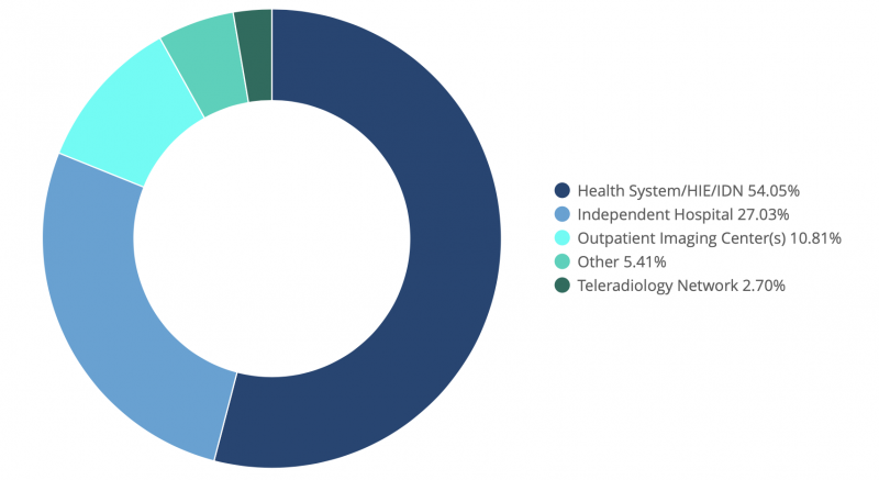
What are Their Top Reasons
for Attending?
Continuing education (CE) and visiting the technical exhibit were
in close contention for the top reason for most respondents attending RSNA this year,
once again demonstrating the show as a destination for scientific and technological
education, advancement and procurement. Of course, one cannot dismiss the popularity of
social and professional networking events — and the many dinners and happy hours that
are sure to be well attended (see below).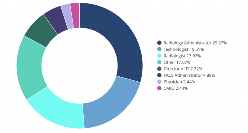
What are
the Top Trends
for RSNA 2018
By aggregating the responses to questions related to continuing
education (CE) topics of interest, active shopping plans for the technical exhibit, and
what new innovations/ technologies and associated applications respondents were most
excited to see at this year’s show we were able to identify five key trends for RSNA
2018 (which we will expand upon in the following sections):
Trend #1: Continued Shift Toward Subspecialty Practice
Once again imaging
modalities are the most popular shopping list item among RSNA attendees this year, with
30 percent of respondents noting that they will be actively shopping for new modalities
— in particular magnetic resonance imaging (MRI) and ultrasound systems — at the show
this year. Following this trend, advances in modality technologies and best practices
lead in terms for CE topics of interest, with 30 percent of respondents planning to
attend sessions related to scientific and technological innovation, dose management and
imaging workflow optimization across a wide array of specific clinical specialty
applications, once again with a heavy emphasis on MRI.
Driven by shifting
reimbursements and growing consumer demand for specialized care services, this trend
highlights the increased focus on subspecialty practice, and subsequently image
acquisition workflow. Consequently, healthcare organizations are seeking to leverage new
technologies to enable more accurate diagnosis, improve clinical outcomes and derive
increased value from their imaging service lines.
Trend #2: Focus on Optimization
Rather Than Replacement
For those who are actively shopping at this year’s show,
analytics/business intelligence (BI) and AI were in contention for second place at the
top of the list, with 21 percent and 20 percent of respondents, respectively. The top
reasons cited for adopting these technologies include:
Conversely, taking a popularity hit from previous years was enterprise imaging systems
and PACS replacements, each appearing on fewer than 9 percent of attendees’ shopping
lists. This is a surprising statistic, given the strong focus on both of these topics at
previous shows.
Combined, these statistics highlight a strong shift toward
optimization of existing systems and workflows and away from large-scale system
replacements, and a demand for evidence-based decision making for both clinical and
operational roles. This results in a greater-than-ever need for interoperability between
vendors — especially up-and-coming BI and AI companies — to support the level of
integration required to deliver on the expectations of this year’s
shoppers.
Trend #3: The Nebulous Potential of AI
It should come as no
surprise that AI was a leading topic for both CE sessions and the technical exhibit. At
a high-level most respondents noted that they were looking to AI to optimize workflow,
improve efficiency and accuracy, and increase satisfaction scores for providers and
patients alike. These are common yet ambiguous answers that continue to reinforce trend
#2 above, however very few respondents offered specific examples when asked what
specific use cases and benefits respondents are hoping AI could offer. This is highly
indicative that AI is still in its healthcare infancy, and that the industry in general
continues to be unsure about what the most immediate and impactful applications of this
emerging technology could be.
With AI appearing in numerous CE sessions and
vendor booths, this year’s show should provide some much-needed education about what AI
is, the role of related machine and deep learning technologies, and what they mean for
healthcare. However, it remains to be seen whether RSNA 2018 will shed some light into
which near-term applications will really take off in the diagnostic imaging realm, and
whether vendors will be able to move the needle in terms of adoption in
2019.
Trend #4: Cloud Confusion
Somewhat unexpectedly, cloud-hosted
solutions demonstrated a lack of interest among active shoppers, appearing on only 7
percent of respondents’ shopping lists, and not appearing at all on lists related to CE
topics of interest or exciting new innovations. This is a surprising result, given the
continued pressure on healthcare organizations to operationalize budgets and reduce
cost, and eludes to lingering uncertainty regarding the technical, operational, or
economic feasibility of vendor-managed infrastructure, systems and data within the
industry at large.
Cloud technology has become a standard data model in many
other industries, and offers several advantages including reduced operational complexity
and capital expense and improved redundancy and security. The lack of adoption to-date
reinforces healthcare’s slow movement to new technologies in general, which is further
exacerbated by the confusion resulting from differing capabilities, pricing models, and
business cases across vendors in the industry. Ultimately, spurring adoption of this
new(er) technology in healthcare will begin with education regarding the capabilities
and benefits of cloud-hosted environments so that IT leaders can become informed
consumers and gain the confidence and trust required to move forward.
Trend #5:
What Didn’t Make the List?
Equally telling to what respondents identified as ‘hot
topics’ is what didn’t make the cut. A couple of the technologies that have been leading
headlines and water-cooler conversations in 2018 include blockchain and consumer
wearables, however they failed to emerge as top-of-mind for respondents when asked what
new and exciting innovations they were hoping to see at the
show.
Blockchain
Most popular for its applications in the financial
industry, blockchain has been lauded for its potential in managing patient identities
and sharing information and images between unaffiliated domains, as well as for the role
it could play in supporting population health, AI and machine learning by aggregating
data and making it accessible for research and algorithm training purposes. However,
very few within the healthcare industry truly understand what blockchain is and how it
works, and more questions than answers exist when it comes to the feasibility of
deploying this resource-intensive technology, which is likely why it failed to make an
appearance.
Consumer Wearables
Innovations like the Apple Watch with the
ability to take ‘anytime, anywhere’ ECG readings continues to be a popular topic of
discussion. While a good idea in theory, care providers have challenged whether the
industry is ready for this type of innovation — citing questions such as how the
resulting information can be meaningfully shared with and used by care providers, what
to do when there is an inevitable influx of false positives to manage, and how will this
fit into existing reimbursement models? These are not trivial concerns, which is why it
is likely too soon for this type of technology to make a meaningful appearance on the
RSNA 2018 agenda.
Conclusion
The trends identified for RSNA 2018 reinforce
that the industry’s top priorities continue to be measurably improving the quality,
scalability and cost of care. Imaging leaders and practitioners alike are cautiously
looking to technology to provide transparency into clinical and business operations,
optimize workflow and resource utilization, and improve interpretation
accuracy.
While the rate of adoption typically lags behind the pace of innovation
in healthcare, it will be interesting to see if this year’s show will provide the
knowledge and confidence needed to diffuse some of the industry’s newer technologies
into clinical practice in 2019.
About the Authors
Jef
Williams is Managing Partner at Paragon Consulting Partners LLC. With decades of
Healthcare IT experience, he has a diverse and creative approach to problem-solving
and solution design. He specializes in assisting organizations with organizational
strategy, operational improvement, software design and selection, and enterprise
imaging deployment initiatives. Williams contributes to the industry at-large as a
member of the AHRA, IHE and by participating in the HIMSS-SIIM working
group.
Laurie Lafleur is a senior imaging consultant at Paragon Consulting
Partners, providing consultative and advisory services for healthcare and technology
organizations. Having worked with care providers and vendors across Canada, the
United States and Europe, she brings over 18 years of relevant experience in
software engineering, product marketing, and business strategy within the Healthcare
IT and imaging informatics industries. She is a member the IHE Radiology Planning
Committee, and serves as a technical advisor on an academic Software &
Electronics Engineering advisory
board.
Reference
1. 2018 Forbes, ‘Theranos?
Whatever. Healthcare startups have raised $15 billion so far this year’,
https://www.forbes.com/sites/matthewherper/2018/07/09/theranos-whatever-healthcare-startups
Published from:
Williams, Jef, and Laurie Lafleur.
"5 Trends to Watch for at RSNA 2018." Imaging Technology News. November 09, 2018.
Accessed November 13, 2018. https://www.itnonline.com/article/5-trends-watch-rsna-2018.

November 7, 2018
Patients want to learn imaging results as soon as possible, from their own doctor
Patients prefer to receive imaging results as
soon as
possible, according to new research published in Radiology. They also want that
information to come from their physician and over the telephone, not through a patient
portal.
“Although
hospitals are implementing [patient portals] at an accelerated rate, there is a lack of
guidance regarding how best to release potentially sensitive information such as
radiologic test results, which fosters variability in hospital
policies,” wrote lead author Matthew S. Davenport, MD, of Michigan Medicine and the
University of Michigan in Ann Arbor, and colleagues. “Some centers may prioritize the
ability of the physician to communicate results before a patient
is able to see them whereas other centers may prioritize the ability of the patient to
view the results without physician oversight.”
Davenport et al. noted that it
sometimes takes physicians one or two weeks to find time to
communicate results to a patient. A patient portal, meanwhile, can allow results to be
shared within three days, but there may not be physician oversight. Either time frame
may increase anxiety on the part of the patient.
The
researchers sought to measure patient preferences regarding the method of communication
and the timing of when imaging results should be communicated. A total of 418 patients
completed a “discrete choice conjoint survey” following an
imaging exam from December 2016 to February 2018. The survey consisted of three
questions regarding patient preferences for receiving results for a potential or known
cancer diagnosis.
Overall, the researchers found that 43
percent of the surveyed patients had experience with an online patient portal. Of those
who had experience utilizing it, 95 percent liked like the process. About 61 percent of
patients said they would use the portal in the future.
“We
found that when given a series of choices in the context of a known or possible cancer
diagnosis, patients preferred to receive imaging results as soon as possible, from their
physician, and over the telephone,” Davenport and
colleagues noted. “In the clinically relevant scenario of immediate release of results
followed by short-term follow-up, an office visit at seven days will be more preferable
than immediate release if immediately released results are
not followed by an office visit within two days or a telephone call within six
days.”
The survey also revealed that most patients did not want to receive their
imaging results before their physician—rather, they wanted the
results at the same time or after their physician received the results. The telephone
was the overall preferred method of communication, followed by an office visit and then
the patient portal. Not surprisingly, those who had prior
experience with a patient portal were less likely to want to wait for their results in
comparison to those who did not have previous experience with such a system.
In
an accompanying editorial, author Ronald L. Arenson, MD, of
the University of California, San Francisco, noted that online portals please most
patients as they are very timely and do not impact a physician’s time. There is one
caveat, however: cancer.
To combat the anxiety around
embargoed receipt of cancer imaging communication, Arenson suggested that physicians
should be able to have their reports embargoed until the physician can review. Another
option would be to give patients the choice between an
immediate release or waiting for the physician to review.
“Our results can be
used to inform local policy and indicate that the embargo period can and possibly should
vary by site, depending on the availability of referring
physicians and their extender staff to contact patients with results,” Davenport et al.
concluded. “Future study into offering patients the ability to customize the use and
length of their specific embargo period may be warranted
because different patients and those in different age groups may have different wishes
with regard to receipt of sensitive imaging results.”
Post-Hurricane Michael Advisory
CMS Imaging, Inc. is continuing to monitor the aftermath of Hurricane Michael and its
effects in Florida.
Our Charleston Call Center is open and accepting service and
part related
phone calls and will continue to operate under normal conditions. For service calls,
please call us at 800.867.1821, email us at service @cmsimaging.com,
or visit our website.
Service in
Florida and other affected areas will return to normal where conditions allow our Field
Service Engineers to commute to
facilities safely. Counties under a Mandatory Evacuation* order from the governor will not be serviced until
that order is lifted. Counties under Voluntary/Phased Evacuations**
orders will be serviced on a case by case basis depending on the conditions of the roads
and the safety of our staff.
All service calls submitted during the evacuations
will be responded to in the order they were
received.
FedEx and UPS delivery services have begun operation in all areas, not
under emergency evacuation orders and deliveries will start today.
Please
contact us at info@cmsimaging.com with any
questions.
Hurricane Advisory - Michael 3
CMS Imaging, Inc. is continuing to monitor Hurricane Michael and has begun to execute our
Storm Contingency Plan along the Florida Panhandle, Big Bend Coast, and surrounding
areas. Hurricane Michael is now a Category 4
storm with maximum sustained winds of 150mph. These winds will also occur well inland
across portions of the Florida Panhandle, southeast Alabama, and southwestern Georgia as
the core of the hurricane moves inland. The
worst of the storm surge is expected later today and tonight with a possible 9 to 14
feet of inundation. Heavy rainfall from Michael could produce flash flooding from the
Florida Panhandle and Big Bend region into
portions of Georgia, the Carolinas, and southeastern Virginia.
Keeping our team
members and their families safe during this storm is our primary concern. In compliance
with the governor of Florida service
operations in those areas affected by the storm have been suspended. CMS Imaging will
continue to monitor the decisions made by the governors of Florida, Alabama, Georgia,
North Carolina, and South Carolina with regards
to the possible suspension of service in subsequent areas.
Our Storm Contingency
Plan is in effect, and we stand prepared to continue our high level of service to our
clients. Our Charleston Call Center is
operating under normal conditions. In the event the Charleston area is impacted by the
effects of the storm, we will switch to our secondary call center. Should our secondary
location also be affected, we have planned a
third off-site call center location to continue to answer and respond to service calls.
All call centers can be reached at our usual phone number, 800.867.1821 or via
email at service@cmsimaging.com.
Additionally, FedEx and
UPS delivery services have suspended pickup and delivery service in some affected areas.
Expect delivery delays in accordance with FedEx and UPS delivery suspension. For more
information on deliveries, please visit
UPS and FedEx.
For those who require
service after the storm, we will begin to dispatch our service engineers to areas
affected by the storm once the federal, state and local
authorities have deemed it safe to return and travel in the areas affected by Hurricane
Michael.
We urge all those in the expected path of Hurricane Michael to monitor
the National
Oceanic and Atmospheric Administration (NOAA) website.
For additional
information on how to prepare for hurricanes, visit the Federal Emergency Management Agency's (FEMA)
website.
The CMS Imaging, Inc. Executive team will meet again tomorrow,
and another Storm Alert will be issued.
Please contact us
at info@cmsimaging.com
with any questions regarding CMS Imaging's Storm Contingency Plan.
Hurricane Advisory - Michael 2
CMS Imaging, Inc. is continuing to monitor
Hurricane
Michael and has begun to execute our Storm Contingency Plan along the Florida Panhandle,
Big Bend coasts and surrounding areas.
Hurricane Michael is now a Category 2 storm and has the potential of making landfall as
a Category 3 hurricane on Wednesday evening.
Keeping our team members and their
families safe during this storm is our
primary concern. Governor Scott of Florida has issued mandatory evacuations along ten
counties, as such service operations in those areas are subject to suspension as the
storm nears.
As the storm proceeds inland,
it has the potential of impacting our service operations throughout inland Florida,
Georgia, and South Carolina. Our Storm Contingency Plan is in effect, and we stand
prepared to continue our high level of service to our
clients. Our Charleston Call Center is operating under normal conditions. In the event
the Charleston area is impacted by the effects of the storm, we will switch to our
secondary call center. Should our secondary
location also be affected, we have planned a third off-site call center location to
continue to answer and respond to service calls. All call centers can be reached at our
usual phone number, 800.867.1821,
email at service@cmsimaging.com, and by
visiting our website.
For those who require
service after the storm, we will begin to dispatch
our service engineers to areas affected by the storm once the federal, state and local
authorities have deemed it safe to return and travel in the areas affected by Hurricane
Michael.
We urge all those in the
expected path of Hurricane Michael to monitor the National Oceanic and Atmospheric
Administration (NOAA) at https://www.noaa.gov.
For additional information on how
to
prepare for hurricanes, visit the Federal Emergency Management Agency's (FEMA) website
at https://www.Ready.gov/hurricanes.
The CMS Imaging, Inc.
Executive team will meet again tomorrow, and another Storm Alert will be issued.
Please contact us at info@cmsimaging.com with any questions
regarding CMS Imaging's Storm
Contingency Plan.
Tropical Storm Advisory - Michael 1
CMS Imaging, Inc. is closely monitoring
Tropical Storm
Michael a storm that is expected to become a hurricane. Michael is now located just east
of Mexico’s Yucatan Peninsula and is
expected to make landfall along the Alabama / Florida Panhandle later this week. Current
forecasts expect Michael to be a Category 2 storm when it impacts the US coastline. The
storm is then expected to swing northeast
and impact Georgia, South Carolina, and North Carolina before heading back out to sea.
It is imperative to us as an organization that all of our clients and team
members remain safe in the affected areas. It is
also our mission to ensure that all of our clients continue to receive the excellent
service to which they are accustomed.
Our teams in both Jacksonville, FL and
Charleston, SC will continue to monitor the
official weather updates from our federal, state and local authorities. Our Service
Management Team and Service Engineers will be presented with an operational plan for
their specific territories once the path of the
storm becomes more definitive.
We have already put in place a contingency plan
for the areas not expected to be directly impacted by this storm. In the event our
Charleston Call Center is directly affected by the
storm, we will automatically switch to an offsite call center to receive and dispatch
our service calls. In the event the offsite location is also affected, we have planned a
tertiary location where our calls will be
answered. All call centers can be reached at our usual phone number, 800.867.1821 via email at service@cmsimaging.com, or our website at
https://www.cmsimaging.com/servicerequest.html.
For those who require service after the storm, we will begin to dispatch our
service engineers to areas affected by the storm once the federal, state and local
authorities have deemed it safe to return and travel
in the areas affected by the storm.
We urge all those in the expected path of
Hurricane Michael to monitor the National Oceanic and Atmospheric Administration (NOAA)
at https://www.noaa.com.
For
additional information on how to prepare for hurricanes, visit the Federal Emergency
Management Agency's (FEMA) website at www.Ready.gov/hurricanes.
The
CMS Imaging, Inc. Executive team will meet again today, and another Storm Alert will be
issued tomorrow.
Please contact us at info@cmsimaging.com with any questions
regarding CMS Imaging's Storm Plan.
CMS Imaging welcomes Steven Oeth to our Team!
CMS Imaging would like to welcome Steven Oeth to the team as an Associate Field Engineer in the Oxford, MS territory. Steven's first day will be October 22, 2018!
CMS Imaging welcomes two new members to our Team!
Please join us in welcoming Rocky Lester to
our Sales
Team! Rocky joins CMS Imaging as a Medical Accounts Manager in lower South Carolina and
Eastern Georgia. Rocky will be taking over for Karen Kadavy as she transitions to
Management role within the CMS Imaging Corporate team. Rocky comes to CMS with many
years of experience selling diagnostic x-ray equipment in the Northeastern United
States. Rocky's first day with CMS will be October 15, 2018.
CMS Imaging is also
welcoming Howard Johnstone to our Service Team! Howard will be joining CMS Imaging as
our new Customer Service Manager. Howard is replacing Tammy Smith as she transitions to
an administrative role with our Project Management Team. Howard's first day will
September 27, 2018.
Shimadzu Medical Systems teams up with Change Healthcare Cardiology™
San Diego—TCT 2018 - September 21,
2018
– Shimadzu Medical Systems USA and Change Healthcare, have entered a
partnership through which Shimadzu will offer Change Healthcare Cardiology™ Hemo, along
with its Trinias line of angiography systems. The announcement was made today at the
annual Transcatheter Cardiovascular Therapeutics (TCT 2018) meeting in San
Diego.
More than 130 million adults in the U.S. are projected to have some form of
cardiovascular disease (CVD) by 2035. The costs associated with caring for CVD are
unsustainable, and reimbursement models are shifting. To support providers looking to
grow their cardiovascular service line and implement technology while maintaining
financial responsibility, augmenting the Trinias line is part of a complete
interventional lab package.
“We are proud to solidify a partnership with Change Healthcare, and extend the
capabilities of their cardiology
hemodynamics solution to achieve a shared initiative of providing healthcare
organizations with solutions that help them meet their operational and quality
initiatives,” said Tim Stevener, Director of Cardiology NA for Shimadzu Medical Systems.
“Our companies are well aligned to provide an unmatched cardiology offering. With this
collaboration, we look forward to a promising relationship with Change
Healthcare.”
“This partnership shows our commitment to continue to work with other industry leaders
to provide options for customers that will help them grow their business while providing
quality care to the populations they serve,” said Aaron Green, SVP and GM, Cardiology,
Change Healthcare. “The combination of the Trinias system and Change Healthcare
Cardiology Hemo provides a unique offering for organizations looking for a complete
solution with mutually exclusive benefits to meet the high demands of a hybrid
interventional lab.”
Trinias is a unique single plane system available in floor or ceiling mounts or as a
Bi-plane mounted system, offering unmatched design movements around the patient. Change
Healthcare Hemo is the integrated hemodynamic monitoring system for cardiology
departments that need to aggregate hemodynamic data, waveforms, and images in one
cardiac patient record. The combined offering is now available through Shimadzu and its
reseller network.
About Shimadzu
Shimadzu Corporation, founded in 1875 in Kyoto, Japan and the parent of Shimadzu Medical
Systems USA (SMS), is a global provider of medical diagnostic equipment including
conventional, interventional and digital X-Ray systems. Shimadzu Medical Systems USA is
headquartered in Torrance California with Sales and Service offices throughout the
United States, the Caribbean and Canada with Direct Operations headquartered out of
Cleveland, OH; Dallas,
TX; and the greater Chicago area. Visit SMS at: www.shimadzu-usa.com or call (800)
228-1429.
Disaster Strikes - What's the plan for your Urgent Care Center?
Arabzadeh, Payman, et al. “Disaster Strikes-What's the Plan for Your Urgent Care Center?” Journal of Urgent Care Medicine, 29 Aug. 2018, www.jucm.com/disaster-strikes-whats-plan-urgent-care-center/.
Urgent message: Urgent care centers exist to help people who need to see a healthcare
professional today. When that need coincides with a natural or manmade disaster, every
location must have a plan of action to ensure any downtime is minimal, staff needs are
met, and the business is able to survive.
Introduction
No region of the country—for that matter, no state, town, neighborhood, or block—is
immune from disabling disasters. Hurricanes, tornadoes, flooding, forest fires,
earthquakes, even lightning strikes don’t care whether yours is a single location owned
by the physician on duty, part of a hospital system, or the flagship of a national
chain. Urgent care centers face the same risks as any other structure, while at the same
time existing for the purpose of helping area residents who need medical care right
away.
The challenge is, how do you ensure you’re prepared not only to protect
your business, but to ensure your staff’s personal and professional needs are met and
that your patients have somewhere to turn no matter what else is going on in the
world?
We invited a handful of true leaders in our industry just that. Here’s
what they had to say.
Do you have an operational
disaster plan specific to your urgent care clinic in place? If so, how often is the
plan reviewed and updated?
ARABZADEH: Davam Urgent
Care does have an operational disaster plan in place, and it’s reviewed and updated at
least biannually. It takes into account multiple forms of potential disasters. We have
every new hire read it prior to coming on board. However, we also communicate with the
staff throughout the year to keep everyone educated and up to date on procedures
necessary in times of potential disasters.
LAMELAS: We have a
well-developed and time-tested plan that addresses each location, operations, corporate,
staffing, etc. We review it yearly, before the start of every hurricane season in
Florida. We also always all sit down at the end of every hurricane season, or after any
actual disaster, to do a postevent analysis. We review what happened, our performance,
and any immediate improvements that might be needed going forward. We learn something
new from every hurricane.
SELLARS: Many of our urgent care
centers are also located in areas vulnerable to hurricanes, so our centers—our entire
business—could lose a great deal if we didn’t have the right disaster plan in place.
Besides direct damage in areas where low elevations and local waterways invite flooding,
we need to be prepared for other consequences including power outages, infrastructure
and property damage, mold, and contaminated water supply. Not many businesses can
withstand being put on hold for very long, so our disaster plan is reviewed at least
annually, sometimes more often depending on the need.
AYERS: We
consult with independent, network, and hospital-affiliated urgent care centers across
the country. The disaster risks, needs, and mitigation strategies vary significantly by
region of the country. In California, it may be earthquakes and wild fires, hurricanes
along the East and Gulf coasts, and blizzards or tornadoes in the Midwest. We advise
every urgent care to have a disaster and business continuity plan in place that’s
appropriate for the risks in their operating area. All new staff members should be
oriented on the plan, annual training should occur on the plan (just prior to the season
of highest risk), and the plan should be reviewed by leadership at least annually for
relevance and detail. It’s also a good idea to engage in disaster simulations—to
actually walk through everyone’s role and the steps to be followed.
Who do you communicate with on a governmental level regarding
curfews, civil disaster plans (eg, a formal state of emergency), and your ability to
open your urgent care clinic?
BEVIS: The director of
the county Emergency Management System is our point of contact in local government
before and during disasters.
ARABZADEH: Constant communication
with local government is key to making sure our community knows whether we are available
to help in a disaster situation. They usually can get out information faster and to a
broader audience then we might be able to. When Hurricane Harvey hit Houston, about an
hour away, we were in touch with our local fire department to let them know whether we
were open or closed. The more people know their options for securing medical needs, the
better off the community will be.
LAMELAS: We do not have
ongoing, direct communication unless the health department, local municipalities, fire
rescue, and police departments reach out to us. Having weathered a few hurricanes since
opening here in south Florida in 2005, they all know we stay open as long as safely
possible, and then try to reopen locations that are safe and have electricity or a
generator as soon as possible. We do, however, keep an up-to-the-minute list of all our
open sites and times on our webpage. Social media and radio are ideal for keeping the
public informed.
SELLARS: One challenge is that phone systems,
the internet, and cell phones may fail or be unreliable during a disaster. Trusted
relationships are important to surviving a disaster situation; those may include our
hospital partners caring for our patients should our centers be closed; local, county,
and state government offices that have certain expertise and resources available in
relieving disaster-related problems beyond our capabilities; and local media and social
media outlets to provide updates to the community on operating hours and status. We also
communicate with our vendors and other business partners as needed, and law enforcement
agencies if necessary.
AYERS: Emergency medical services,
hospital emergency rooms, and primary care providers should be aware of the urgent care
center’s availability in case of a natural disaster. Developing local relationships and
keeping others appraised of the urgent care’s operating status can result in referrals
to urgent care when physician offices are unavailable, and knowing that ambulatory,
nonemergent patients can walk in to urgent care can open capacity for first responders
and hospital EDs to focus on more acute needs in the community. Because urgent care
benefits from patient referrals, it’s always a good idea to develop relationships with
community providers, regardless of whether there’s a disaster.
How do you decide who to schedule if you do stay
open?
SELLARS: Our chief operating officer serves as the Incident Commander
during any disaster situation and works with the owners, partners, and other members of
the administrative team to initiate and coordinate all aspects of the Emergency
Preparedness Plan.
BEVIS: First we decide if it is safe for
patients and our staff to be open. From there, our regional directors work with staff to
determine who is able to cover shifts.
ARABZEDAH: Yes, in the
event of a disaster, scheduling is based on who is available to come in, depending on
the severity of the disaster. Those who have not been affected, and can safely come to
work, come help if possible. We have an in-house communication program that we used to
keep in constant communication with the staff during normal working hours, as well as in
disaster situations. The leadership team continually monitors and discusses
implementation of these plans on a disaster-by-disaster
basis.
LAMELAS: We are not open during the hurricane, and we try
to close 24-36 hours before the event, depending on the storm, since the track is not as
well established until then. I make that final call with input from my executive team
and weather reports. As a physician, I always want the patients to come first, without
risking the safety of our staff, and like to say, “We are here for our patients and
because of our patients.”
Who can (or should) be on
call if needed?
LAMELAS: From an urgent care
operations perspective, that is the single most difficult part of dealing with any
hurricane here in south Florida. School and daycare closings that force parents to stay
home, people being evacuated from their homes or having difficulty traveling because of
downed trees or power lines, as well as big and small things you would have never
thought of, affect staffing. We try to determine which staff live in an evacuation zone,
then create a list of staff and providers willing to take calls and be available. Lots
of people step up during these difficult times and perform above and beyond what is
expected. Others do not. It is always good to hope for the best, but be prepared by
staffing extra employees, calling them in advance of their shifts, and having on-call
staff and back-up plans. The most important thing is to communicate clear and concise
messaging with staff before and after the hurricane.
SELLARS:
While no staff members are expected to take any action that may endanger his/her life,
all clinical staff members are considered essential and are expected to report to work
to provide medical care to the community. Disaster on-call teams are established to
provide additional coverage at high-volume clinics.
What if staff members have been affected directly by the
disaster?
SELLARS: Adequate staffing is critical to
meet increased healthcare demands during and after a disaster. However, staff may become
both victims and responders when a disaster occurs. When this happens, staff member
availability may be impacted for the first several days following a disaster. At Premier
Health, we make every effort to assist staff members with their own personal disaster
planning by incorporating preparedness efforts into our overall
communications.
LAMELAS: We try to be supportive of our staff
and keep open lines of communication, emails, group text, phone contact, radios, etc. on
an ongoing basis. If employees have been personally affected, we try and shift them
around between open sites so they can keep their hours/pay, give bonuses and extra
personal time for outstanding performance, and even have been very charitable to them in
times of need.
AYERS: Larger urgent care providers often have
an employee assistance fund, to which employees can apply for support when facing
individual hardship. Employees can donate to help their peers and there’s a review
process to determine which requests are funded. Smaller operators will provide direct
contributions, paycheck advances, or collections among employees. Also, its’ very common
for employees to be able to donate their unused paid time off to colleagues in
need.
And what’s the procedure for letting staff and
patients know when your location has been affected—by physical damage or power
outages, for example? Do you refer patients elsewhere on your website or building
signage?
ARABZADEH: We use our social media network
(Facebook, Google Plus, Instagram, website, etc.) to get information directly to our
patients. We do our best to find out which alternate locations, whether it be hospitals,
emergency rooms, or other urgent cares, are open and to pass the information along to
our patients until we can get our location back up and
running.
BEVIS: In 2017 we had an automobile crash into our
front lobby. Our immediate priority was to ensure the safety and health of our patients
and staff. Fortunately, nobody was seriously hurt. We did, however, have to immediately
close the clinic. We called 911, and local fire and police departments arrived quickly
to secure the area. We announced on signage, social media, and phone messages that we
would close temporarily. Patients were referred to neighboring
clinics.
LAMELAS: MD Now locations have been affected in
different ways over the years—loss of power, window and building damage, floods, etc.,
so we have had single and multisite closures. In addition to our website, social media,
and front-door signage, we use our office phone outgoing message to refer patients to
our open sites or the ED. We email our corporate accounts and call patients proactively
to reschedule appointments. After the storm, we do status call three to five times a day
between management teams and corporate, and also group texts. It’s helpful to have a few
different methods of communication available.
SELLARS: We
utilize an Emergency Notification Call Tree to communicate with staff members. It’s
distributed to all staff and posted at all worksite locations. Communication may be in
the form of text, phone, email, social media, or through a dedicated emergency message
phone line with recorded message updates. The Incident Commander coordinates messages
with senior managers, regional administrators, and other associates that may include
hospital command centers, media, insurance carriers, utility services, law enforcement,
and others. If necessary, patients may be referred to affiliated entities through press
releases, on websites, social media channels, LED signs, and temporary signs. In short,
it’s important to have a plan to communicate before, during, and after a disaster
through alternate channels, if necessary.
What have
you learned from prior natural disasters that would be relevant to operating your
urgent care clinic?
SELLARS: We’ve experienced
catastrophic flooding, civil unrest, and three hurricanes in markets where we operate
urgent care centers in the past year. All of these situations required the activation of
our Emergency Preparedness Plan. The most important lesson we’ve learned is to have a
well-developed disaster plan in place, and to clearly and emphatically communicate that
plan to all staff members before a disaster occurs. Advanced planning is critical to
ensure the plan is implemented and executed effectively. That said, what may look good
on paper doesn’t always apply in a real-life situation, so it’s extremely important to
spend time not just preparing but to regroup after the disaster to make necessary
adjustments.
ARABZADEH: We have had the luxury of having “only”
one major disaster in our tenure here, but it was a horrible hurricane that resulted in
catastrophic flooding. From that we have learned to always expect the unexpected. There
was no way anyone could have prepared for the amount of damage and destruction that
occurred. Our biggest takeaway was the fact that constant communication with your staff
and outside emergency services will ensure the quickest and safest possible road to
recovery.
LAMELAS: Four very important lessons: 1) Frequent
communication is the single most important factor. 2) Planning, preparing, and training
far in advance of any storm or event is essential, as is reviewing the disaster plan
annually. 3) Selecting a responsible and available executive team and
managers/supervisors, with a clear chain of command is a requirement. 4) Order extra
medications—both in-house meds and dispensed prescription meds—plus certain vaccines,
such as tetanus, and address proper storage issues. Other essential medical supplies,
water, and even toilet paper have to be stocked, as well, upon increased probability of
hurricane impact by weather models.
Not all valuable lessons come from within urgent
care, by the way. I actually am very impressed and would like to model our clinics to
perform as well as the most time-tested hurricane-impacted business in south Florida:
believe it or not, Publix Supermarkets. They have weathered these storms for over 87
years, and they’re among the last to close and first to reopen. As urgent care
operators, we need to think outside the box (or typical healthcare) and consider all
industry “best practices.”
AYERS: You have to balance what’s
right for the employees, patients, and what’s needed in the community. One client
decided to keep its centers open during a blizzard (an unusual occurrence in their
temperate climate), notifying its primary care and hospital ED partners that the urgent
care would be available. Generators were connected to power the centers, employees were
housed in hotels near the centers, overtime labor costs were incurred, and referral
providers were notified urgent care would be available. But because virtually nobody in
the community was leaving home, the centers only saw three or four patients during the
days of the storm and as a result, the urgent care lost its shirt in its service to the
community, not to mention frustrating employees who were kept away from home and
family.
What role can urgent care operators play in
helping communities start to rebuild?
BEVIS: When an
F5 tornado hit Lutts, TN in 2015 we sent providers, nurses, and staff with supplies to
render emergency medical care and relief supplies. This was all possible because of our
close proximity to this community. More recently, with hurricanes Irma and Harvey, we
established drop-off locations for much needed supplies at each of our clinics. We
consolidated donations at our Waynesboro, TN headquarters and loaded a 50-foot trailer
to each impacted state. Announcements were made through social media and
email.
LAMELAS: Urgent care centers play an increasingly
important role in the healthcare infrastructure and local medical community. At MD Now,
we have been involved in community events, local outreach, charitable events, and
contributions in the U.S., but especially in south Florida and the local communities we
serve. We also support the Red Cross and others in relief efforts here and abroad as we
did during the earthquake in Haiti by donating resources, money, and medical
supplies.
It goes beyond our patients; urgent care is becoming more and more a
respected and essential “medical safety net” that hospitals, private physicians and
medical practices, and our communities cannot live without. Having said that, the urgent
care industry as a whole is still not well integrated and connected into the U.S.
healthcare infrastructure. I believe we need to remedy that through continuing outreach
efforts, both as an industry and individually, to further develop the formal integration
of our clinics and specialty into that existing infrastructure.
SELLARS: We can and should play an important role in disaster recovery.
After serious, damaging floods hit the Baton Rouge area last year, we initiated a
campaign called We’re Here for You to help meet the community’s healthcare needs during
the time of crisis. Despite some clinics experiencing flooding of more than 4 feet of
water, our volunteer team of administrative, clinical, and medical providers worked in
the 100⁰ postflood heat to feed, treat, and comfort displaced residents. With many cell
towers down, we informed residents about clinic openings and closings, operational
hours, tetanus shot information, and school/road closures on Facebook. Additionally, our
medical leadership team appeared on local TV and radio to direct residents where to seek
treatment for flood-related and other health issues, as well as to provide tips for safe
flood cleanup.
ARABZADEH: As an urgent care operator, the more
you can help and give back to your community, the more successful and sought-after you
will become. In the aftermath of Hurricane Harvey, our biggest goal was giving back to
the community. We encouraged others to do the same by posting on our social media
networks that we were taking in donations for those who had been affected by the
flooding. Most recently, we partnered up with UCA for the weekend of service, in which
we provided free medical assistance to those affected by the flooding. We are also
running a flu shot drive, with the proceeds going to those affected by the flooding and
hurricane damage.
Coming together as a family to help the community and to open the urgent care back up as
soon as possible, without causing extra risk to anyone, can truly be the cornerstone of
recovery during and after a disaster.
CMS Imaging, Inc. Post-Hurricane
Advisory
September 17, 2018
CMS Imaging, Inc. is continuing to monitor the
aftermath
of Hurricane Florence and its effects in North Carolina and South Carolina.
Our
North Charleston Call Center is open and accepting service and part related phone calls
and will continue to operate under normal conditions. For service calls, please call us
at 800.867.1821, email us at service@cmsimaging.com, or visit our website
Service in
South Carolina will return to normal where conditions allow our Field Service Engineers
to commute to facilities safely.
Service in North Carolina will return to normal
in areas not under the governor's evacuation orders and where the facility is
acceptable. Counties under a Mandatory Evacuation*
order from the governor will not be serviced until that order is listed. Counties under
Special Condition (Mixed) Evacuation** orders will be
serviced on a case by case basis depending on the conditions of the roads and the safety
of our staff.
All service calls submitted during the evacuations will be
responded to in the order they were received.
FedEx and UPS delivery services
have begun operation in all areas, not under emergency evacuation orders and deliveries
will start today.
Please contact us at info@cmsimaging.com with any
questions.
*NC Counties under Mandatory Evacuation (as of 9/17/18 at 9:00 am)
**NC Counties under Special Condition (Mixed) Evacuation (as of 9/17/18 at 9:00 am)
CMS Imaging, Inc. Hurricane Advisory
3
September 12, 2018
CMS Imaging, Inc. is continuing to monitor
Hurricane
Florence and has begun to execute our Storm Contingency Plan in Georgia, South Carolina,
and North Carolina.
The current expected track of the storm is indicating that
the storm will linger in North Carolina and South Carolina and possibly Georgia
throughout the weekend. If this is the case, please expect continued delays in service
until federal, state and local authorities deem it safe to return to those areas.
Keeping our team members and their families safe during this storm is our
primary concern. Evacuations have begun to along the coastal communities of North
Carolina and South Carolina, and our North Charleston office is closed.
Our
Charleston Call Center has shifted to our usual weekend service call center which will
continue to serve our customers, not in the impacted areas.
Our weekend call
center will operate normally unless the path of the storm places that call center in
jeopardy. If that should occur, our tertiary call center location will take over and
continue to answer and respond to service calls. All call centers can be reached at our
usual phone number, 800.867.1821 or via email at service@cmsimaging.com.
Additionally, FedEx and UPS delivery services have suspended pickup and delivery
service in some areas of the evacuation zones. Expect delivery delays in accordance with
FedEx and UPS delivery suspension. For more information on deliveries, please visit UPS and FedEx.
For those who require service after the storm, we will begin to dispatch our
service engineers to areas affected by the storm once the federal, state and local
authorities have deemed it safe to return and travel in the areas affected by Hurricane
Florence.
We urge all those in the expected path of Hurricane Florence to monitor
the National Oceanic and Atmospheric Administration (NOAA) at https://www.noaa.com.
For additional
information on how to prepare for hurricanes, visit the Federal Emergency Management
Agency's (FEMA) website at www.Ready.gov/hurricanes.
Please contact us at info@cmsimaging.com with any questions
regarding CMS Imaging's Storm Contingency Plan.
CMS Imaging, Inc. Hurricane Advisory
2
September 11, 2018
CMS Imaging, Inc. is continuing to monitor
Hurricane
Florence and has begun to execute our Storm Contingency Plan in the Carolinas. Hurricane
Florence is now a Category 4 storm with winds exceeding 140mph and is expected to make
landfall along the North Carolina and South Carolina coast during the latter part of the
week.
Keeping our team members and their families safe during this storm is our
primary concern. The governors of North Carolina and South Carolina have issued
mandatory evacuations along their respective coasts.
In compliance with the
governor of South Carolina, CMS Imaging, Inc will be closing our North Charleston
corporate offices beginning on Wednesday, September 12, 2018. Our Storm Contingency Plan
is in effect and we stand prepared to continue our high level of service to our clients.
Our Charleston Call Center is continuing to operate with normal service today and will
switch calls to our secondary call center beginning after business hours today. In the
event our secondary location is also affected, we have planned a third off-site call
center location to continue to answer and respond to service calls. All call centers can
be reached at our normal phone number, 800.867.1821 or via
email at service@cmsimaging.com.
For those who require service
after the storm, we will begin to dispatch our service engineers to areas affected by
the storm once the federal, state and local authorities have deemed it safe to return
and travel in the areas affected by Hurricane Florence.
Additionally, FedEx and
UPS delivery services have suspended pickup and delivery service in the evacuation
zones. Expect delivery delays in accordance with FedEx and UPS delivery suspension. For
more information on deliveries, please visit the UPS and FedEx
websites.
We urge all those in the expected path of Hurricane Florence to
monitor the National Oceanic and Atmospheric Administration (NOAA) at https://www.noaa.com.
For
additional information on how to prepare for hurricanes, visit the Federal Emergency
Management Agency's (FEMA) website at https://www.Ready.gov/hurricanes.
The CMS Imaging, Inc.
Executive team will meet again tomorrow and another Storm Alert will be issued.
Please contact us at info@cmsimaging.com with any questions
regarding CMS Imaging's Storm Contingency Plan.
CMS Imaging, Inc. Hurricane Advisory
1
September 10, 2018
CMS Imaging, Inc. is closely monitoring
Hurricane
Florence, a category 2 storm with maximum sustained winds of 105 mph, which is currently
located in the southeast of Bermuda. Florence is now heading toward the east coast of
the United States before posing a serious threat to South Carolina, North Carolina, and
Virginia beginning Thursday of this week.
It is imperative to us as an
organization, that all of our clients and team members remain safe in the affected
areas. It is also our mission to ensure that all of our clients continue to receive the
excellent service to which they are accustomed.
Our teams in both Jacksonville,
FL and Charleston, SC will continue to monitor the official weather updates from our
federal, state and local authorities. Our Service Management Team and Service Engineers
will be presented with an operational plan for their specific territories once the path
of the storm becomes more definitive.
We have already put in place a contingency
plan for the areas not expected to be directly impacted by Hurricane Florence. In the
event our Charleston Call Center is directly affected by the storm, we will
automatically switch to an offsite call center to receive and dispatch our service
calls. In the event the offsite location is also affected, we have planned a tertiary
location where our calls will be answered. All call centers can be reached at our normal
phone number, 800.867.1821 via email at service@cmsimaging.com, or our website at
https://www.cmsimaging.com/servicerequest.html.
For
those who require service after the storm, we will begin to dispatch our service
engineers to areas affected by the storm once the federal, state and local authorities
have deemed it safe to return and travel in the areas affected by Hurricane
Florence.
We urge all those in the expected path of Hurricane Florence to monitor
the National Oceanic and Atmospheric Administration (NOAA) at https://www.noaa.com.
For
additional information on how to prepare for hurricanes, visit the Federal Emergency
Management Agency's (FEMA) website at https://www.ready.gov/hurricanes.
The CMS Imaging, Inc.
Executive team will meet again today and another Storm Alert will be issued tomorrow.
Please contact us at info@cmsimaging.com with any questions
regarding CMS Imaging's Storm Plan.
Scanning the dead: Orange County votes to buy CT machine to examine deaths
The Orange County (Orlando-Kissimmee-Sanford,
Florida)
commissioners approved the request of Dr. Joshua Stephany, the chief medical examiner in
Orange and Osceola counties to purchase a computerized tomography scanner (CT scanner)
on Tuesday, July 31, 2018, from CMS Imaging, Inc. CT Scanners are standard in hospitals
but are unusual in US autopsy rooms. The Neusoft Neuviz 16 Essence CT scanner will not
replace traditional autopsies, but the digital images may assist county and city
officials in solving crimes. These digital autopsies will allow medical examiners an
alternative to preserving the deceased's dignity and also adhere to religious tenets
with regards to traditional autopsies.
Read the Full Article*
*
will take you to an external website

July 23, 2018
Shimadzu Medical Systems USA is proud to announce the release of the RADspeed fit.
Torrance, CA — July 23, 2018 - Shimadzu
Medical Systems
USA, a subsidiary of Shimadzu Corporation, is proud to announce the release of the
RADspeed fit.
Shimadzu Medical Systems USA has launched a new radiographic
table system called the RADspeed fit, a general radiography table system offering higher
image quality than any other compact integrated system in its class.
As the
newest U.S. based product in the RADspeed series, the RADspeed fit with its integrated
tube stand, is an outstanding radiography system that offers a cost-effective balance of
functionality to support a wide range of general rad applications, such as chest,
abdomen, or extremities. The fit can perform emergency examinations while providing the
same easy operability and extensive functionality for reducing exposure levels as
developed for other products in the series.
The system generator is
integrated into the patient table and with easy removal of the tabletop, servicing the
unit is a snap making it ideal as an entry level digital radiography (DR) system.
Practically priced, the RADspeed fit is equipped with a digital X-ray detector (FPD) and
makes the perfect digital X-ray system for Urgent Care and Clinical Centers.
The RADspeed fit will be on display at the AHRA in Orlando, FL (July 23-25, 2018) in
booth #404. About Shimadzu
Shimadzu Corporation, founded in 1875 in Kyoto,
Japan and the parent of Shimadzu Medical Systems USA (SMS), is a global provider of
medical diagnostic equipment including conventional, interventional and digital X-Ray
systems. Shimadzu Medical Systems USA is headquartered in Torrance California with Sales
and Service offices throughout the United States, the Caribbean and Canada with Direct
Operations headquartered out of Cleveland, OH; Dallas, TX; and the greater Chicago area.
Visit SMS at: www.shimadzu-usa.com or call (800)
228-1429
Change Healthcare Selects Avreo, Inc., as Radiology Information Systems (RIS) Partner
Change Healthcare’s Imaging, Workflow &
Care
Solutions business, and Avreo, Inc., a developer of radiology workflow solutions,
announced a partnership today during AHRA 2018, the association for medical imaging
management’s Annual Meeting and Exposition in Orlando, Fla.
“We are thrilled
to solidify a partnership with Change Healthcare and extend the capabilities of
interWORKS to achieve a shared initiative of providing healthcare organizations with the
most positive user experience possible,” said John Sloan, CEO of Avreo. “Our companies
are well matched and possess the same core values, standards, and work ethic. With this
collaboration, we look forward to a promising relationship with Change
Healthcare.”
InterWORKS is a single application/single database Radiology
Workflow Solution (RWS) that includes RIS, PACS, Dictation, Transcription, and an auto
report delivery system that encompasses the features a practice or hospital needs to
operate efficiently.
“This partnership provides an option for our customers
that are looking for a RIS-based workflow in conjunction with our Radiology PACS.” said
Todd Johnson, Executive Director, Radiology Product Development, Change Healthcare. “At
Change Healthcare’s, we are committed to providing our customers with the solutions that
we know can meet their needs.”
AHRA 2018 runs from July 22-25 in Orlando. The
four-day event, with more than 150 exhibitors and over 70 educational sessions, is
expected to attract more than 1,000 medical imaging leaders.
Avreo® provides EHR for Radiology, RIS, PACS, Dictation, Transcription, and Report
Distribution services in a single application, single database system. This innovative
approach provides healthcare professionals with user-friendly access to information in
one streamlined system. Avreo offers solutions tailored to a variety of healthcare
organizations including hospitals, imaging centers, radiology groups, and
orthopedics.
Avreo is a privately held company headquartered in Charleston,
SC. Learn more at www.changehealthcare.com.
Change Healthcare (CHC) is a leading provider of software and analytics, network solutions and technology-enabled services that optimize communications, payments and actionable insights designed to enable smarter healthcare. By leveraging its Intelligent Healthcare Network™, which includes the single largest financial and administrative network in the United States healthcare system, industry stakeholders are able to increase revenue, improve efficiency, reduce costs, increase cash flow and more effectively manage complex workflows. Learn more at www.changehealthcare.com.
Canon Products Win 2018 iF Design Awards
Canon Inc. has announced that several Canon
product
designs were recognized by iF International Forum Design GmbH with prestigious 2018 iF
Design Awards.
Established in 1953, the iF DesignAwards are recognized,
worldwide, as one of the most prestigious awards within the field of design, with
outstanding industrial designs chosen from all over the world each year. This year 6,402
entries from 54 countries and regions were judged by internationally active design
experts across seven disciplines: product, packaging, communication, service design,
architecture, interior architecture, and professional concept.
Canon has now
won the iF Design Award for 24 consecutive years. Encouraged by the recognition of the
Company’s design excellence, Canon will continue striving to realize products that
combine high levels of performance and design.
The following Canon products
have been recognized as winners of the iF Design Award 2018:
First-ever colour X-ray on a human performed

PARIS: New Zealand scientists have performed
the
first-ever 3-D, colour X-ray on a human, using a technique that promises to improve the
field of medical diagnostics, said Europe’s CERN physics lab which contributed imaging
technology.
The new device, based on the traditional black-and-white X-ray, incorporates
particle-tracking technology developed for CERN’s Large Hadron Collider, which in 2012
discovered the elusive Higgs Boson particle.
“This colour X-ray imaging technique could produce clearer and more accurate
pictures and help doctors give their patients more accurate diagnoses,” said a CERN
statement.
The CERN technology, dubbed Medipix, works like a camera detecting
and counting individual sub-atomic particles as they collide with pixels while its
shutter is open.
This allows for high-resolution, high-contrast
pictures.
The machine’s “small pixels and accurate energy resolution meant
that this new imaging tool is able to get images that no other imaging tool can
achieve,” said developer Phil Butler of the University of
Canterbury.
According to the CERN, the images very clearly show the
difference between bone, muscle and cartilage, but also the position and size of
cancerous tumours, for example.

The technology is being commercialised by New Zealand company MARS Bioimaging, linked to the universities of Otago and Canterbury which helped develop it.
Konica Minolta Introduces AeroRemote Insights: Interactive Analytic and Business Intelligence Reporting for Radiology
WAYNE, N.J., May 05, 2018 (GLOBE NEWSWIRE) --
Konica
Minolta Healthcare Americas, Inc., a recognized leader in medical imaging systems and
healthcare IT, announces the introduction of AeroRemote™ Insights, a cloud-based,
business intelligence and analytics solution that delivers detailed information on asset
utilization, department workflow and efficiency, system health and more. AeroRemote
Insights provides daily performance data in intuitive visual formats that managers can
utilize to optimize department performance and manage digital radiography assets. This
new service also enables managers to act on urgent situations immediately and respond to
usage trends intelligently.
“Representing a new era of proactive monitoring solutions, AeroRemote Insights
delivers the information department managers need in today’s competitive healthcare
environment to make better informed decisions, sooner,” says Steven Eisner, Senior
Product Manager at Konica Minolta. “It is a great addition to Konica Minolta
Healthcare’s top-rated customer service by delivering valuable, interactive and
actionable data wherever and whenever needed.”
AeroRemote Insights provides vital information on productivity, user performance and
AeroDR system health at a glance. Easy to use data analytics are available from any
computer or mobile device and will enable departments to improve workflow, accuracy and
uptime. AeroRemote Insights represents Konica Minolta’s continuing investment in IoT,
machine learning and artificial intelligence by creating analytic tools that increase
the value of conventional hardware and software solutions.
“With this innovative approach to proactively monitoring our systems, we have
revolutionized traditional service delivery,” says Kevin Chlopecki, Vice President of
Service Operations. “We address our customers’ common productivity and performance
challenges, allowing healthcare providers to stay committed to patient care. As a
result, our service teams can gather data, predict appropriate responses and solve
common issues—often before our customer knows of a situation. Our brand, our vision and
our obsession are providing a seamless customer experience. AeroRemote Insights offers a
cost-effective, competitive differentiator to Konica Minolta customers,” says Chlopecki.
AeroRemote Insights is available for most Konica Minolta AeroDR® Systems as part of
Konica Minolta’s premium Blue Moon service plans or as a stand-alone subscription.
About Konica Minolta Healthcare Americas, Inc.
Konica Minolta Healthcare is a world-class provider and market leader in medical
diagnostic imaging and healthcare information technology. With over 75 years of endless
innovation, Konica Minolta is globally recognized as a leader providing cutting-edge
technologies and comprehensive support aimed at providing real solutions to meet
customer's needs and helping make better decisions sooner. Konica Minolta Healthcare
Americas, Inc., headquartered in Wayne, NJ, is a unit of Konica Minolta, Inc.
(TSE:4902). For more information on Konica Minolta Healthcare Americas, Inc., please
visit
www.konicaminolta.com/medicalusa.

May 25, 2018
Initial Clinical Results of Konica Minolta Healthcare’s Dynamic Digital Radiography Presented at ATS 2018 Annual Meeting
WAYNE, N.J., May 24, 2018 (GLOBE NEWSWIRE) -- Konica Minolta Healthcare Americas, Inc.,
announced that two clinical studies utilizing Dynamic Digital Radiography (DDR), the
company’s innovative X-ray technology under development, were presented
at the American Thoracic Society (ATS) 2018 Annual Meeting this week. Dynamic Digital
Radiography is a new modality that utilizes conventional X-ray images and Konica
Minolta's proprietary software platform to create X-ray in Motion. DDR
is the most significant advancement in X-ray since digital radiography was introduced.
X-ray in Motion of the chest provides a visual display of the dynamic interactions
of lung, muscle, bone, heart and nerve, not captured by either a conventional static
X-ray or the common pulmonary function test (PFT). This allows clinicians
to visualize and quantify physiological changes in anatomical structures during the
complete respiratory cycle. Previously, respiration could only be radiologically
assessed using fluoroscopy, which involves higher levels of radiation exposure.
X-ray in Motion may better enable clinicians to evaluate chest and lung function in
patients with pulmonary diseases, such as chronic obstructive pulmonary disease (COPD),
asthma and lung cancer.
At the ATS meeting, the study, A New Technology: The Dynamic Image of a Forced
Breath Compared to a Tidal Breath Uncovers a Physiological Phenomenon in COPD, was
presented, demonstrating a correlation to the severity of COPD and diaphragm
excursion during forced and tidal breathing using DDR. The research concluded that DDR
may be a clinically relevant option to assess COPD severity in the acute setting and for
patients unable to perform PFTs. This study was conducted by
the Division of Pulmonary, Critical Care and Sleep Medicine, Department of Medicine, and
Department of Radiology from Mount Sinai (New York, NY).
The research study, Evaluation of Pulmonary Function Using Dynamic Chest
Radiographs: The Change Rate in Lung Area Due to Respiratory Motion Reflects Air
Trapping in COPD, was also presented. The researchers investigated DDR as an alternative
for the evaluation of pulmonary function in COPD patients by examining the change rate
in lung area due to respiratory motion in patients with COPD and in subjects with normal
pulmonary function. The study found that DDR is a viable alternative
indicator for air trapping in COPD. Air trapping is the abnormal retention of air in the
lungs making it difficult to exhale completely. This study was performed by the
Departments of Respiratory Medicine, Thoracic, Cardiovascular and General
Surgery, and College of Medical, Pharmaceutical & Health Sciences at Kanazawa
University (Kanazawa, Japan).
Digital Radiography, or X-ray, is a primary diagnostic technology that is widely
available throughout the world. Konica Minolta is investing in the development new
clinical applications and tools for existing technology, such as digital
radiography, that can deliver more information to clinicians than previously attainable
while supporting the reduction of healthcare expenditures. Using readily available X-ray
systems, DDR can provide incremental value for diagnosis, ongoing
disease management, preoperative planning and post-operative assessment, without
subjecting the patient to multiple and often more expensive tests.
Dynamic Digital Radiography represents ongoing research and development at Konica
Minolta. It is not cleared by the FDA for commercial distribution or available for sale
in the United States.
About Konica Minolta Healthcare Americas, Inc.
Konica Minolta Healthcare is a world-class provider and market leader in medical
diagnostic imaging and healthcare information technology. With over 75 years of endless
innovation, Konica Minolta is globally recognized as a leader providing
cutting-edge technologies and comprehensive support aimed at providing real solutions to
meet customer's needs and helping make better decisions sooner. Konica Minolta
Healthcare Americas, Inc., headquartered in Wayne, NJ, is a unit of Konica
Minolta, Inc. (TSE:4902). For more information on Konica Minolta Healthcare Americas,
Inc., please visit
www.konicaminolta.com/medicalusa.

May 25, 2018
An update to Our Privacy Policy and our Terms and Conditions
CMS Imaging, Inc. has updated our Privacy
Policy and
Terms and Conditions as of May 24,2018. These changes were made in preparation for the
European Union's new data privacy law, the General Data Protection Regulation (GDPR).
With these updates , CMS Imaging, Inc. reaffirms our commitment to safeguarding the
personal data of our contacts and anyone who visits our website. We embrace privacy by
design, which means we will continue to build features with privacy
considered alongside innovation and functionality.
We encourage you to take the time to review our revised Privacy Policy and Terms and
Conditions. By continuing to use CMS Imaging, Inc's website on or after May 24, 2018 you
acknowledge our updated Privacy Policy and Terms and Conditions.
As CMS Imaging, Inc continues to grow and evolves, we will continue to focus on
strengthening and improving our privacy policies and tools, for the benefit of our
contacts and website visitors.
See our new Privacy Policy at www.cmsimaging.com/privacy.html
See our new Terms and Conditions at www.cmsimaging.com/terms.html
For more information visit contact us at: privacy@cmsimaging.com

May 14, 2018
CMS Imaging welcomes Christopher Ryan
CMS Imaging welcomes Christopher Ryan to the
Team!
Chris will be our new Medical Accounts Manager in South Florida, reporting to JB
Glass. Chris's first day will be Monday, May 14, 2018.
Please join us in welcoming Chris!
How much are patients paying out of pocket for advanced imaging services?
Patients are often required to pay high
out-of-pocket
costs for advanced imaging services, especially when out of their network, according to
a new study published in the Journal of the American College of Radiology. The authors
suggested
that radiologists should communicate these costs to patients to help prevent “surprise
billing.”
“To interact more effectively with their patients, health systems and policymakers,
radiologists should become more familiar with the various factors influencing their
patients’ out-of-pocket expenses for imaging examinations in their
local private insurance market places,” wrote lead author Andrew B. Rosenkrantz, MD,
MPA, of the department of radiology at NYU Langone Medical Center in New York. “With
greater awareness of patients’ out-of-pocket cost responsibilities,
radiologists may be more judicious in recommending additional or follow-up imaging,
knowing that those recommendations frequently translate into high costs borne
substantially, and sometimes completely, by their patients.”
The authors, wanting to take a deep dive into these out-of-pocket costs, turned to
extensive data from the 2017 CMS Health Insurance Marketplace Benefits and Cost Sharing
Public Use file. Overall, 48 percent of health plans required
coinsurance for advanced imaging services, more than 9 percent required copays and 8
percent required both. More than 34 percent of plans required no coinsurance or copay.
For out-of-network advanced imaging services, more than 91 percent
of plans required coinsurance, 0.1 percent required copayments and 1 percent required
both. More than 7 percent of those plans required no coinsurance or copay.
The study also found that, when deductibles are present, patients were responsible
for more than 27 percent through coinsurance in network and more than 47 percent out of
network. And when deductibles were present, the average copays
were $319 in network and $630 out of network. With no deductible present, the average
coinsurance burden for patients was 99.9 percent.
“Overall, patients’ out-of-pocket cost responsibilities for advanced imaging are
quite variable and can be particularly high in many circumstances because advanced
imaging most commonly requires coinsurance from patients,” the authors
wrote.
Rosenkrantz et al. also looked at state-level variation in the average coinsurance
burden for patients both in and out of network. For example, the average in-network
patient coinsurance was more than 33 percent in New Jersey and just
1.4 percent in Alabama.
“There was a tendency for out-of-network coinsurance responsibilities to be higher
in lower income states,” the authors wrote. “This observation highlights how variation
in the benefit design of available plans at the regional level
can potentially create disproportionate cost burdens on financially disadvantaged
patients. Such economic disparities could limit vulnerable populations’ access to
imaging services.”
In this era of value-based care, knowing more about these statistics and using that
information to educate patients is especially important. “To optimally guide individual
patients in an era of shared decision making in an environment
of imaging stewardship, radiologists may need to integrate the findings of studies they
interpret with further awareness of individual patients’ insurance coverage and
financial means to afford recommended testing,” Rosenkrantz and colleagues
concluded. “Through such understanding, radiologists may be able to help patients become
better informed upfront about types of imaging examinations and potential alternative
sites of service, thus reducing their risk for large surprise
bills.”

April 19, 2018
Konica Minolta Healthcare Introduces KDR Primary to Meet the Digital Radiography Needs of Office-based Imaging Providers
WAYNE, N.J., April 19, 2018 (GLOBE NEWSWIRE) -- Designed to meet the
needs of office-based X-ray imaging, such as orthopedic groups, family practices and
urgent care centers, Konica Minolta Healthcare Americas, Inc. introduced
the KDR Primary Digital Radiography System at the American Association of Orthopedic
Executives (AAOE) 2018 Annual Conference. KDR Primary delivers advanced digital X-ray
capabilities and exceptional functionality in a compact footprint.
It is the ideal solution for practices and clinics that want superior image quality and
versatile imaging options yet have a limited amount of space.
Building on the success of the KDR Advanced U-arm System, KDR Primary helps optimize
workflow, expedite the diagnostic process and increase staff efficiency all while
elevating the patient experience. Equipped with a 17 x 17 inch detector,
KDR Primary captures high resolution images in seconds, providing detailed bone and soft
tissue visualization from a single exposure. Practices and clinics can enhance patient
satisfaction by providing rapid diagnostic results without the
need to schedule additional appointments or refer elsewhere.
KDR Primary has a wide range of motion to enable all required imaging views. The
swivel arm rotates 135 degrees in both directions and moves 39 inches vertically while
the detector tilts 45 degrees in two directions to accommodate patients
who are standing, sitting, lying on a table or confined to a wheelchair. The space
saving design provides staff with the freedom to maneuver the system for true exam
flexibility that can translate to exceptional patient care and potentially
help reduce retakes due to incorrect patient positioning or movement. It can be
installed in a standard room with an 8 ft. ceiling. Options include a weight-bearing
stand and pediatric imaging package.
“At Konica Minolta, we are focused on answering our customer’s clinical needs by
delivering new innovations in imaging technology such as KDR Primary that allow for more
personalized patient care,” says Kirsten Doerfert, Sr. Vice President
of Marketing, Konica Minolta Healthcare. “The KDR Primary fulfills an imaging need for
which our customers have been asking for a solution and provides the same superior
imaging, reliability and performance inherent in all our DR systems.”
About Konica Minolta Healthcare Americas, Inc.
Konica Minolta Healthcare is a world-class provider and market
leader in medical diagnostic imaging and healthcare information technology. With over 75
years of endless innovation, Konica Minolta is globally recognized
as a leader providing cutting-edge technologies and comprehensive support aimed at
providing real solutions to meet customer's needs and helping make better decisions
sooner. Konica Minolta Healthcare Americas, Inc., headquartered in Wayne,
NJ, is a unit of Konica Minolta, Inc. (TSE:4902). For more information on Konica Minolta
Healthcare Americas, Inc., please visit www.konicaminolta.com/medicalusa.

April 4, 2018
Konica Minolta announces the addition of the AeroDR HD 10” x 12” and 17” x 17” Flat Panel Detectors
Konica Minolta is pleased to announce the
addition of the
AeroDR HD 10” x 12” and 17” x 17” Flat Panel Detectors. These two new sizes join the
AeroDR HD 14” x 17” detector to round out Konica Minolta’s AeroDR product line. AeroDR
HD is ideal
for use in a wider range of environments, including orthopedic, general radiography,
ICU, emergency departments and operating rooms.
The AeroDR HD 100 μm sampling pitch (pixel size) delivers outstanding resolution
that is among the highest in the world. It allows orthopedists to capture clearer images
of fine structures, such as finger trabecula, contributing to ease
of diagnosis.
AeroDR HD is the only detector to offer both High Definition and High Dynamic Range
modes.
The AeroDR HD features the most advanced technology in our lightest and toughest enclosure:
For more information visit our webpage for AeroDR HD Flat Panel Detectors HERE

March 28, 2018
The Centers for Medicare and Medicaid Services Qualifies MedCurrent OrderWise® Clinical Decision Support Mechanism (CDSM) for Appropriate Use of Advanced Diagnostic Imaging
Toronto, ON (PRWEB) April 04,
2018 -
MedCurrent, a global leader in innovative healthcare IT solutions, has received full
qualification by the Centers for Medicare and Medicaid Services (CMS) for its clinical
decision support
mechanism (CDSM), OrderWise®. Through this qualification, OrderWise® is recognized for
establishing technology and processes to support healthcare providers improve the
quality of patient care, manage costs and meet regulatory requirements
for the appropriate use of advanced imaging.
Starting January 1, 2020, Medicare outpatient reimbursement for advanced diagnostic
imaging procedures (MR, CT, NM, PET) will be at risk if ordering providers do not
consult clinical decision support (CDS) as initially set forth in the
Protecting Access to Medicare Act (PAMA, 2014). To meet the requirements of this
mandate, only qualified CDSMs and Appropriate Use Criteria (AUC) developed by qualified
provider-led entities (qPLEs) may be used.
“We are pleased to receive full qualification by attesting to the standards set
forth by CMS. This is an opportunity to support healthcare providers in the US make
evidence-based decisions for their patients,” says John Adziovsky, Chief
Executive Officer (CEO), MedCurrent. “Our new status represents a significant milestone
on our journey to continuous innovation of our CDS offering and enabling clients to
improve outcomes.”
MedCurrent’s OrderWise® guides providers through clinical decision pathways to help
them order the right imaging test for their patient. The solution is complemented by
robust Analytics and Authoring Studio modules to support quality
improvement initiatives and system customization. In the US, OrderWise® integrates
imaging AUC from Intermountain Healthcare, Sage Evidence-based Medicine & Practice
Institute (SEMPI), National Comprehensive Cancer Network (NCCN) and
the American College of Cardiology (ACC). Internationally, OrderWise® now runs with the
eighth edition of iRefer: Making the best use of clinical radiology, the most up-to-date
referral guidance from The Royal College of Radiologists (RCR).
For more information on the AUC program and qualified CDSMs, please visit the
Centers for Medicare and Medicaid Services (CMS).
About MedCurrent
MedCurrent is a physician-founded Clinical Decision Support (CDS) company focused
on improving the quality of care and managing health system costs through our innovative
and scalable solution, OrderWise. Our solution enhances the clinical
decision-making process with real-time, evidence-based guidelines integrated at the
point of care to improve health and healthcare delivery. Deep healthcare experience,
superior technology and business agility make MedCurrent a global leader
in CDS solutions. More information about MedCurrent is available at https://www.medcurrent.com.
Further information:
Hasan Dharamshi, VP Professional Services
MedCurrent
1.855.279.3380 ext. 503
hasan.dharamshi(at)medcurrent(dot)com

March 28, 2018
CMS Imaging welcomes Chip Bindhamer as the new Medical Accounts Manager in Georgia
North Charleston, SC — March 28,
2018 -
CMS Imaging, Inc. welcomes Chip Bindhamer to the team! Following the promotion of JB
Glass to Regional Sales Director, Chip will be transitioning into JB's previous role
after his onboarding.
Chip's first day will be April 2, 2018.
March 15, 2018
CMS Imaging, Inc and Maury Regional Medical Center bring advanced x-ray and fluoroscopy technology to Middle Tennessee
NORTH CHARLESTON, SC - Maury Regional Medical
Center in
Columbia, TN has announced the beginning of usage of a Shimadzu Sonialvision G4
purchased from CMS Imaging, Inc. The Sonialvision G4 brings the "Best in Class"
performance of a multi-functional
universal Radiography / Fluoroscopy system to Middle Tennessee.
"The system offers physicians advanced imaging technology while minimizing X-Ray
dose levels and can accommodate patients weighing up to 700 pounds. Currently, we are
the only provider in Middle Tennessee to offer this system," said
Maury Regional Health CEO Alan Watson.
"CMS Imaging is proud to partner with Maury Regional to bring this exciting and
advanced technology to the people of Middle Tennessee. The Shimadzu Sonialvision G4 will
provide the physicians of Maury Regional ultra-high resolution
x-ray and fluoroscopic images while reducing the x-ray dose," said CMS Imaging, Inc
Senior Strategic Accounts Manager John Lillie.
The Sonialvision G4 is designed to consider all users in a diversity of
situations, making it ideal for a wide variety of examinations, such as Orthopedics,
General Radiography Studies, Gastrointestinal Studies, Endoscopy, Urology,
and Angiography.
Shimadzu's Sonialvision G4 offers a consistently high image quality. It's
best-in-class, 139 µm ultra-small pixel pitch FPD and SUREengine Advance,
state-of-the-art digital image processing technology ensure the highest image quality
ever.
Additionally, it achieves comprehensive dose reduction as a total system, equipped
with various functions to reduce radiation exposure levels effectively such as removable
grid, ideal Grid-controlled Pulsed fluoroscopy, new collimator
with Multi-Beam Hardening (MBH) filters, Virtual Collimation and above-mentioned high
quality imaging ability itself will also contribute to even further dose reduction
possibility while keeping good output image quality.
To learn more about the Shimadzu Sonialvision G4, please visit our website at
https://www.cmsimaging.com/sonialvisiong4.html or contact us at info@cmsimaging.com.
About CMS Imaging, Inc:
CMS Imaging, Inc. is the premier healthcare solutions provider specializing in the
sales and service of diagnostic medical imaging equipment. Founded in 1987 in
Charleston, SC as an independent service organization, CMS Imaging has
expanded its product line to include MRI, CT, Digital X-Ray, and Advanced Fluoroscopic
systems. Software solutions including Avreo, Authpal by Availity, PACS Harmony, and
MedCurrent's Clinical Decision Support (CDS) are also available to
enable customers to display, manage, and store imaging data.
About Maury Regional Medical Center:
Serving more than 200,000 people across southern Middle Tennessee, Maury Regional
Medical Center, a 255-bed facility, is the largest medical center between Nashville and
Huntsville and had been evaluated by outside organization and
compared to some of the most respected medical centers in the country. For 2018, Maury
Regional Medical Center was ranked number one on Tennessee for overall hospital care and
overall surgical care in the area of medical excellence by CareChex.com
- an information service of Quantros, Inc. The medical center offers a wide range of
advanced services including an accredited heart program, neonatal intensive care and
cancer center. In addition to Maury Regional Medical Center, Maury
Regional Health includes Marshall Medical Center in Lewisburg, Wayne Medical Center in
Waynesboro, Lewis Health Care in Hohenwald, Maury Regional Medical Group physician
practices, as well as additional facilities across southern Middle
Tennessee.
About Shimadzu Corporation:
Shimadzu Corporation has remained committed to commercializing cutting-edge
technology and providing it to customers in a wide array of industries for more than 140
years. Our brand statement, "Excellence in Science", reflects our
desire and attitude to diligently respond to customers' requirements by offering
superior, world-class technologies indispensable for analytical and measuring
instruments, medical systems, aircraft equipment and industrial machinery in the
area of human health, safety and security of society and advancement of industry. In the
ever-changing landscape of challenges of society, Shimadzu aims to partner with
customers to meet their needs with unique technologies and solutions.
Phil Reichner
843.300.4924
preichner@cmsimaging.com
A Case Study on Automating Prior Authorizations - Presented by Linda D'Amore and Mohammed Ahmed
Six industry leaders, including the AMA,
recently
announced collaborating to improve the prior authorization process. In the statement,
they said “Prior authorization approvals can be burdensome for health care
professionals, hospitals,
health insurance providers, and patients because the processes vary and can be
repetitive. Streamlining approval processes will enhance patient access to timely,
appropriate care and minimize potential disruptions.”
Nowhere is that truer than in independent radiology, where prior authorizations are
required for the majority of procedures. While the industry works to enact wider change,
several innovative practices and solution providers are actively
working to find a better way to manage prior authorizations in radiology. Some have even
implemented fully market capable solutions.
This session will profile one of those fully implemented solutions as a case study
and explore the lessons learned from that journey for others considering this type of
initiative. Hosted by RBMA, this session will explore best practices
as well as lessons learned and remaining challenges for both health IT solution
providers and the radiology organizations seeking a solution. Join us for this panel
interview format discussion of innovation, implementation, and automation
in radiology prior authorizations.
This webinar is complimentary for RBMA members but registration is required here.
About the Speakers
Linda D'Amore is a health care project consultant and an accomplished senior leader
with over 35 years of health care experience in both private practice and hospitals.
She has a strong business development background with a broad range
of strategic, operational and financial service skills. Her passions are leading
organizations through change, service line development, and creating a customer
service environment.
Linda holds a Master of Business Administration from Webster University, a
bachelor's in health administration from the Medical University of South Carolina
and is a Registered Radiologic Technologist.
Mohammed Ahmed is VP of authorizations sales enablement for Availity Provider
Solutions. He manages business development for Availity’s prior authorization
solution and the AuthPal product. He has more than 18 years of experience in health
care and is a serial entrepreneur focused on understanding end-user pain points and
developing high quality, cost efficient solutions.
Mohammed is the former CEO of FORE Support Services, which solved the radiology
prior authorization pain point via automation. Fore Support Services was acquired by
Availity in 2016 and now is integrated in the provider solutions
portfolio.
CMS Imaging, Inc donates portable ultrasounds to local animal charities
March 12, 2018
NORTH CHARLESTON,
SC
- CMS Imaging, Inc. announces the donation of Konica Minolta Sonimage™ P3 portable
ultrasounds to The Avian Medical Clinic, an operating division of The Center for Birds
of Prey located
in Awendaw, SC; the Charleston Animal Society located in North Charleston, SC; and
Pethelpers located in Charleston, SC.
The Sonimage™ P3 portable ultrasounds will assist the veterinarians and medical
staffs at the respective facilities help diagnose the causes of pain, swelling, and
infection in the animal's internal organs. It is also used to help
guide biopsies, diagnose heart conditions, and assess damage after a heart attack.
Ultrasound uses high-frequency sound waves to capture live images from the inside of the
body and is safe, noninvasive, and does not use ionizing radiation.
The Konica Minolta Sonimage™ P3 is a true portable ultrasound machine that gives
medical personnel the ability to do more for patients where and when they need it most –
at the point-of-care. With its small footprint and weighing less
than a pound, this handheld device can accelerate and improve interventions and
decision-making time.
About CMS Imaging, Inc.:
CMS Imaging, Inc. is the premier healthcare solutions provider specializing in the
sales and service of diagnostic medical imaging equipment. Founded in 1987 in
Charleston, SC as an independent service organization, CMS Imaging has
expanded its product line to include MRI, CT, Digital X-Ray, and Advanced Fluoroscopic
systems. Software solutions including Avreo, Authpal by Availity, PACS Harmony, and
MedCurrent's Clinical Decision Support (CDS) are also available
to enable customers to display, manage, and store imaging data.
About The Center for Birds of Prey:
The Center for Birds of Prey is a 501(c)3 organization that continues to lead and
participate in groundbreaking scientific research on avian genetics and environmental
hazards. A “citizen science” approach to a number of initiatives
allows the public to become active contributors to wildlife conservation and to raise
public awareness of vital ecological issues. Citizen-science programs carry the added
bonus of raising public awareness about ecological issues, educating
the public about species of concern and their associated habitats, and allowing the
public to become actively engaged supporters of wildlife conservation. Visit them here:
http://www.thecenterforbirdsofprey.org/
About the Charleston Animal Society:
Since 1874, Charleston Animal Society’s mission has been the prevention of cruelty
to animals. Thanks to supporters like you, we are able to touch the lives of 20,000
animals every year and work every day to make Charleston not only
a No Kill Community but also a humane community that is safe for families, including
pets. Our Vision is one where all healthy and treatable animals are saved. It’s a vision
where all people and animals are treated with respect and kindness.
And it envisions a world where cruelty is not tolerated. Visit them here: https://www.charlestonanimalsociety.org/
About Pethelpers:
Pet Helpers was founded in 1978 by Carol Linville, now President of Pet Helpers,
after she learned that 8,000 pets were being euthanized each year at local shelters. It
began as a weekly “adopt a pet” column. More than 30 years later,
that column has grown into Pet Helpers Adoption Center and Veterinary Clinic, one of the
foremost animal rescue organizations in South Carolina. Pet Helpers has slowly evolved
into a widely recognized and innovative shelter that offers
caring solutions to the serious problems created by pet overpopulation. Visit them here:
https://pethelpers.org/
Phil Reichner
843.300.4924
preichner@cmsimaging.com

March 7, 2018
High-resolution brain imaging provides clues about memory loss in older adults
As we get older, it's not uncommon to experience "senior moments," in which we
forget where we parked our car or call our children by the wrong names. And we may
wonder: Are these memory lapses a normal part of aging, or do they signal
the early stages of a severe disorder such as Alzheimer's disease? Currently, there's no
good way to tell.
University of California, Irvine-led researchers, however, have found that
high-resolution functional magnetic resonance imaging of the brain can be used to show
some of the underlying causes of differences in memory proficiency between
older and younger adults.
The study, which appears today in the journal Neuron, involved 20 young adults (ages
18 to 31) and 20 cognitively healthy older adults (ages 64 to 89). The participants were
asked to perform two kinds of tasks while undergoing fMRI
scanning -- an object memory task and a location memory task. Because fMRI looks at the
dynamics of blood flow in the brain, investigators were able to determine which parts of
the brain the subjects were using for each activity.
In the first task, participants viewed pictures of everyday objects and were then
asked to distinguish them from new pictures. "Some of the images were identical to ones
they'd seen before, some were brand-new and others were similar
to ones they'd seen earlier -- we may have changed the color or the size," said Michael
Yassa, director of UCI's Center for the Neurobiology of Learning & Memory and the
study's senior author. "We call these tricky items the 'lures.'
And we found that older adults struggle with them. They're much more likely than younger
adults to think they've seen those lures before."
The second task was nearly the same but required subjects to determine whether the
location of objects had been altered. Here, older adults fared quite a bit better than
in the prior task.
"This suggests that not all memory changes equally with aging," said lead author
Zachariah Reagh, who participated in the study as a graduate student at UCI and is now a
postdoctoral fellow at UC Davis. "Object memory is far more vulnerable
than spatial, or location, memory -- at least in the early stages." Other research has
shown that problems with spatial memory and navigation do manifest as individuals
progress toward Alzheimer's disease.
Importantly, by scanning the subjects' brains while they underwent these tests, the
scientists were able to establish a cerebral mechanism for that deficit in object
memory.
They found that it was linked to a loss of signaling in a part of the brain called
the anterolateral entorhinal cortex. This area is already known to mediate communication
between the hippocampus, where information is first encoded,
and the rest of the neocortex, which plays a role in long-term storage. It's also an
area severely affected in people with Alzheimer's disease.
"The loss of fMRI signal means there is less blood flow to the region, but we
believe the underlying basis for this loss has to do with the fact that the structural
integrity of that part of the brain is changing," Yassa said. "One
of the things we know about Alzheimer's disease is that this region of the brain is one
of the very first to exhibit a key hallmark of the disease, deposition of
neurofibrillary tangles."
In contrast, the researchers did not find age-related differences in another area of
the brain connected to memory, the posteromedial entorhinal cortex. They demonstrated
that this region plays a role in spatial memory, which was not
significantly impaired in the older subjects.
"This suggests that the brain aging process is selective," Yassa said. "Our findings
are not a reflection of general brain aging but rather of specific neural changes that
are linked to specific problems in object but not spatial memory."
To determine whether this type of fMRI scan could eventually be used as a tool for
early diagnosis, the researchers plan to expand their work to a sample of 150 older
adults who will be followed over time. They will also be conducting
positron emission tomography, or PET, scans to look for amyloid and tau pathology in
their brains as they age.
"We hope this comprehensive imaging and cognitive testing will enable us to figure
out whether the deficits we saw in the current study are indicative of what is later to
come in some of these individuals," Yassa said.
"Our results, as well as similar results from other labs, point to a need for
carefully designed tasks and paradigms that can reveal different functions in key areas
of the brain and different vulnerabilities to the aging process,"
Reagh added.
Story Source:
Materials provided by University of California - Irvine. Note: Content may be edited
for style and length.
Journal Reference:
Zachariah M. Reagh et al. Functional Imbalance of Anterolateral Entorhinal Cortex
and Hippocampal Dentate/CA3 Underlies Age-Related Object Pattern Separation Deficits.
Neuron, 2018 DOI: 10.1016/j.neuron.2018.01.039
University of California - Irvine. (2018, March 7). High-resolution brain imaging provides clues about memory loss in older adults: UCI-led study reveals potential tool for early dementia diagnosis. ScienceDaily. Retrieved March 9, 2018 from https://www.sciencedaily.com/releases/2018/03/180307132005.htm
CMS Imaging welcomes Robert James
North Charleston, SC — February 27,
2018
- CMS Imaging, Inc. welcomes Robert James to the team!
Robert will be a Field Service Engineer serving the Pensacola, FL area. Robert's
first day will be March 26, 2018.

February 20, 2018
SIR 2018 March 17 - 22 / Los Angeles, California - Los Angeles Convention Center
SIR 2018 is designed to meet the educational
needs of
practicing interventional radiologists, IRs in-training, diagnostic radiologists, and
other physicians with a special interest in IR—and is the only venue offering IR
education designed
specifically for clinical associates. The meeting also offers a supportive and highly
interactive introduction to IR for medical students who may be considering the specialty
as their focus.
Be sure to visit the Shimadzu Booth (Booth #441) at the upcoming SIR 2018
conference.
*Offer applies to only those participants within CMS Imaging’s business territory
and during SIR 2018.

Februay 7, 2018
RSNA 2017 Highlights Latest Trends in Digital Radiography - Feature article in Imaging Technology News, January/February Issue
By: Jeff Zagoudis
February 7,
2018
- If there was a theme to digital radiography (DR) advances in 2017, it might have been
“upgrade now or be left behind.” The first of the year saw the inception of the federal
Consolidated
Appropriations Act (CAA) of 2016, which among its many provisions imposed a significant
reimbursement penalty (20 percent) on Medicare claims for imaging exams done with screen
film X-ray systems. As of this year, the law adds a 7 percent
reduction for computed radiography (CR) exams that increases to 10 percent in 2023. The
penalties are meant to further encourage providers and hospitals to modernize their
imaging inventory as healthcare as a whole looks to become more
efficient while maintaining good patient outcomes.
These dual goals of increased efficiency and better patient outcomes have driven
many of the recent major advances in DR technology, including improved image quality at
lower radiation doses and workflow efficiencies to increase patient
throughput. These trends were fully evident in the DR offerings at the 2017 Radiological
Society of North America (RSNA) annual meeting in November.
Ultimately, any DR system is only as effective as its detector technology — the
backbone and opportunity in digital radiography. Government and industry forces are
pushing providers toward DR because it generates a higher-quality image
than CR or film systems. It does so at a significantly lower radiation dose than its
predecessors, balancing the equation that connects diagnostic certainty and patient
safety. Vendors continue to push the envelope of how to maximize image
quality while maintaining low dose, and several offered new solutions at RSNA 2017.
Konica Minolta Healthcare Americas, creator of the AeroDR high definition (HD) panels, introduced a
brand-new image processing software for its CS-7 platform. The software,
dubbed Realism, provides clarity and detail beyond conventional processing, according to
Konica Minolta, by independently processing bone and soft tissue data, and enhancing
sharpness and contrast to improve visualization of traditionally
hard-to-see structures.
Realism is included on the newest detector panels in Konica Minolta’s lineup, a 10 x
12-inch and a 17 x 17-inch AeroDR HD panel. The smaller panel is engineered for the
neonatal intensive care unit (NICU) and/or imaging fine structures
such as the extremities; the 17 x 17 is for larger anatomical areas such as the chest
and abdomen. All AeroDR HD panels offer the option to switch between High Definition
(HD) and High Dynamic Range (HDR) imaging. HD provides images with
a 100 micron pixel size for a high level of detail, while HDR imaging aggregates the
data from four pixels for a wider range of grays, providing a smooth image that allows
clinicians to detect subtle differences in soft tissue.
Just before RSNA, Canon launched the 10 Series wireless
digital detector panels, including the CXDI-710C,
CXDI-810C and CXDI-410C. The panels are the lightest in Canon’s arsenal,
coming in a full 2 pounds lighter than the previous generation, according to the
company. The lightweight and the new ergonomic handgrips carved directly into the
detector make the panel extremely easy to carry around.
For the complete article please visit the Imaging Technology News website.
Frost & Sullivan Acclaims Shimadzu for Delivering a Product Line of Versatile and Innovative Radiography Solutions
Shimadzu’s products utilize
advanced robotic
capabilities and next-generation digital imaging technologies
Based on its recent analysis of the general radiography market, Frost &
Sullivan recognizes Shimadzu Corporation with the 2017 Global Frost & Sullivan Award
for Product Line Strategy Leadership. Constant innovations and upgrades
to Shimadzu’s radiography product portfolio have ensured that the products stay relevant
and in high demand in an evolving radiography market. Its comprehensive product range
enhances automation, efficiency, image quality, and overall
clinical value by enabling flexible positioning, reduced radiation dose, and versatility
in viewing bone and soft tissue images.
Shimadzu has the entire range of general radiography solutions, from fixed
ceiling-mounted to floor-mounted systems and mobile X-ray solutions, with or without one
to two arms. These are all available all in entry-level to high-end
configurations. In 2016, it introduced new technologies such as digital multi-slice
tomography with flexible positioning to offer a view of oblique cross sections of the
spine and hip joints. Other products include:
RADspeed Pro
EDGE and
MobileDaRt Evolution MX7 have already been adopted by more than 100 hospitals worldwide
within a year of their introduction.
“In response to customer demand, Shimadzu introduced a sophisticated,
remote-controlled, auto-positioning feature in the RADspeed Pro EDGE package. This
feature helps the radiographer effortlessly position the tube and fine-tune this
positioning to improve patient safety and reduce radiation dose,” said Frost &
Sullivan Consultant Poornima Srinivasan. “Similarly, it incorporated removable grids to
lower radiation exposure during pediatric imaging, and a dose area
product meter on the collimator to measure the dose of radiation the patient is exposed
to.”
Shimadzu’s products have proven their utility in guiding hospitals towards the era
of value-based healthcare. They achieve that by incorporating a host of novel features
and technologies.
“Shimadzu has leveraged the expertise of key
opinion
leaders (KOLs), designed training and educational programs, expanded geographically, and
made the most of cross-selling opportunities in line with its business goals,” noted Ms.
Srinivasan.
“Additionally, it has partnered with universities to optimize the existing systems and
develop further innovations and efficiencies. Shimadzu’s discussions with international
channels, and focus on gathering input from healthcare professionals
have played a huge role in its development of unique products and provision of a rich
customer experience.”
Each year, Frost & Sullivan presents this award to the company that has
developed a comprehensive product line that caters to the breadth of the market it
serves. The award recognizes the extent to which the product line meets
customer base demands, the overall impact it has in terms of customer value, as well as
increased market share.
Frost & Sullivan Best Practices awards recognize companies in a variety of
regional and global markets for demonstrating outstanding achievement and superior
performance in areas such as leadership, technological innovation, customer
service, and strategic product development. Industry analysts compare market
participants and measure performance through in-depth interviews, analysis, and
extensive secondary research to identify best practices in the industry.
About Shimadzu Corporation
Shimadzu Corporation has remained committed to commercializing cutting-edge
technology and providing it to customers in a wide array of industries for more than 140
years. Our brand statement, "Excellence in Science", reflects our
desire and attitude to diligently respond to customers' requirements by offering
superior, world-class technologies indispensable for analytical and measuring
instruments, medical systems, aircraft equipment and industrial machinery in
the area of human health, safety and security of society and advancement of industry. In
the ever-changing landscape of challenges of society, Shimadzu aims to partner with
customers to meet their needs with unique technologies and solutions.
About Frost & Sullivan
Frost & Sullivan, the Growth Partnership Company, works in collaboration with
clients to leverage visionary innovation that addresses the global challenges and
related growth opportunities that will make or break today's market
participants. For more than 50 years, we have been developing growth strategies for the
global 1000, emerging businesses, the public sector, and the investment community.
Contact us: Start the discussion.
Ana Arriaga
P: 210.247.3823
F: 210.348.1003
E: ana.arriaga@frost.com

February 2, 2018
CMS Imaging welcomes Josue Rodriguez and Alex Adzhigirey
North Charleston, SC — February 27,
2018
- CMS Imaging, Inc. welcomes Josue Rodriguez and Alex Adzhigirey to the team!
Josue will be a Field Service Engineer serving the Wilmington, NC area. Josue hails
from the seaside town of Mayaguez in western Puerto Rico. He is married to the lovely
Jaqaira and is the father of two beautiful daughters, Kiara and
Marina.
Alex will be part of the team serving the central North Carolina area. Alex was born
in Eastern Europe but has lived most of his life in Massachusetts and the Carolina. He
is married to the lovely Anna and the father of four beautiful
daughters and two awesome sons.
Welcome to the CMS Family Josue and Alex!
The first day for both will be Monday, January 16, 2018.
Winter Storm Advisory 1: Wednesday, January 3, 2018
CMS Imaging, Inc. is closely monitoring Winter
Storm
Grayson, a major cold weather event settling on the East Coast from Florida through
Maine. Sub-freezing temperatures, freezing rain, and snow are causing major delays and
closures on the
roads.
It is imperative to us as an organization, that all of our clients and team members
remain safe in the affected areas. It is also our mission to ensure that all of our
clients continue to receive the excellent service to which they
are accustomed.
Our teams in both Jacksonville, FL and Charleston, SC will continue to monitor the
official weather updates from our federal, state and local authorities. Our Service
Management Team and Service Engineers will be presented with an
operational plan for their specific territories once the path of the storm becomes more
definitive.
Our Charleston Call Center has switched to an offsite call center to receive and
dispatch our service calls. In the event the offsite location is also affected, we have
planned a tertiary location where our calls will be answered.
All call centers can be reached at our normal phone number, 800.867.1821 or via email at
service@cmsimaging.com.
For those who require service during the winter storm, we will begin to dispatch our
service engineers to areas affected by the storm once the federal, state and local
authorities have deemed it safe to travel in the areas affected.
We urge all those in the areas affected to monitor the National Weather Center at
www.weather.gov.
The CMS Imaging, Inc. Executive team will meet again today to determine our plans
for tomorrow, January 4, 2018.
Please contact us at info@cmsimaging.com with any questions regarding CMS Imaging's
Winter Storm Plan.

December 25, 2017
Shimadzu Medical Systems USA receives highest rating in MD Buyline Reports - December 25, 2017
TORRANCE, Calif., December 25,
2017 -
Healthcare research firm MD Buyline released its Q4 2017 Market Intelligence Brief™ for
Q4 2017 to its hospital and vendor subscribers. This exclusive survey is based on
nationwide,
direct-user feedback from hundreds of healthcare providers who rely on MD Buyline’s
healthcare market research to guide their critical decision-making in budgeting,
planning, selecting and acquiring medical equipment and technology.
Suppliers are rated on a scale from 1 to 10 in six categories: 1) system
performance, 2) system reliability, 3) installation and implementation, 4) applications
training, 5) service response time and 6) service repair quality. Based
solely on hospital user feedback, the Market Intelligence Brief™ is an unvarnished study
of user opinion that helps MD Buyline's Research and Analysis team to map trending in
both end-user feedback and ratings of suppliers and technologies.
Shimadzu Medical Systems USA has received highest rankings in their class
for overall customer satisfaction in Portable Radiographic Systems & Radiography
and Fluoroscopy for Q4 2017.
Shimadzu's Portable Radiographic family includes: MobileDaRt Evolution MX7 Version
and MobileArt Evolution MX7 Version. The Radiography and Fluoroscopy product line
include: FLUOROspeed and SONIALVISION G4. Shimadzu Medical Systems
can offer multiple DR solutions based on your preference.
About Shimadzu
Shimadzu Corporation, founded in 1875 in Kyoto, Japan and the parent of Shimadzu
Medical Systems USA (SMS), is a global provider of medical diagnostic equipment
including conventional, interventional and digital X-Ray systems. Shimadzu
Medical Systems USA is headquartered in Torrance California USA with Sales and Service
offices throughout the United States, the Caribbean and Canada with Direct Operations
headquartered out of Dallas, Texas, and the greater Chicago area.
Visit SMS at: www.shimadzu-usa.comor call (800) 228-1429
About MD Buyline
Healthcare providers recognize MD Buyline as the leading strategic sourcing company
serving hospitals and vendors today. Guiding hospitals in their critical decision-making
in budgeting, planning, selecting and acquiring medical equipment
and technologies, our expert analysts operate with transparency and trust and help
clients target cost-reduction opportunities for purchased services, consumables, capital
and IT acquisitions. For more than 30 years, hospitals have relied
on MD Buyline’s data and experienced analysts to guide them in their most important
purchasing decisions.
For more information, go to www.mdbuyline.com.
*MD Buyline Technology Report, Q4 2017
CMS Imaging welcomes Brian Bohager - December 7, 2017
North Charleston, SC — December 7,
2017
- CMS Imaging, Inc. welcomes Brian Bohager to the team! Brian will be a Senior Field
Service Engineer working in the Jacksonville Florida Area. Brian comes to us with over
13 years
experience as a service engineer. Brian proudly served in the United States Marine Corp
for 7 years.
Welcome to the CMS Family Brian!
Brian's first day will be Monday, December 11, 2017.
RSNA 2017 - Day 3; November 28, 2017
Chicago, IL — November 28,
2017 - Come
visit our partners Konica Minolta (South Hall Booth 1919) and Del Medical (South Hall
Booth 1929) as they update some of their most trusted products and introduce us to new
tchnology.
See below for photos.
Konica Minolta KDR™ Advanced U-Arm – Advanced design innovations to optimize
workflow, increase staff productivity and improve outcomes. Konica Minolta REALISM™ -
Our new Image processing software. See exceptional bone detail and
elevated contrast of soft tissue for uncompromised clarity. Konica Minolta Exa™
Enterprise Imaging – Access to images across the enterprise. Anytime, anywhere,
securely.
Del Medical introduces updates to the FMT18 and the OTC 18, introducing digital
heads to these most trusted products.
Shimadzu Medical System USA releases MobileDaRt Evolution MX8 Version; November 27, 2017
TORRANCE, Calif., Nov. 27, 2017 -
Shimadzu
Medical Systems USA, a subsidiary of Shimadzu Corporation, is proud to announce the
release of the MobileDaRt Evolution MX8 version.
Shimadzu released the next generation of MobileDaRt Evolution systems, called the
MX8 version. The MX8 is digital mobile X-ray system featuring Shimadzu's first
telescoping support column for even greater drivability with improved
forward visibility. Also featured on the MX8 is a large completely flat monitor screen,
a lockable Flat Panel Detector (FPD) housing compartment and an extensive selection of
options with overall improved operability.
The newly redesigned MobileDaRt Evolution MX8 version was developed to provide
users with multifaceted support based on Shimadzu's extensive technology and track
record cultivated thus far. A telescoping X-ray tube support column
and other changes provide dramatically improved forward visibility during travel, while
maintaining the outstanding drivability that has always been highly regarded by users.
Furthermore, the design also helps improve the efficiency of
hospital rounds, such as by offering a wireless hand switch and other new optional
features, by improving the convenience of the monitor on the mobile unit, the area
around the collimator, and FPD housing compartment, and by using a longer
X-ray focal length to make radiography easier for examining patients on high beds.
Shimadzu is currently promoting a concept referred to as "DR Neutral," which is
based on offering a combination of digital radiography (DR), that is matched to the
operations performed within the hospital, and Shimadzu's radiography
systems. The MobileDaRt Evolution MX8 version can also be used with the newest and
lightest weight FPDs from Canon.
About Shimadzu
Shimadzu Corporation, founded in 1875 in Kyoto, Japan and the parent of Shimadzu
Medical Systems USA (SMS), is a global provider of medical diagnostic equipment
including conventional, interventional and digital X-Ray systems. Shimadzu
Medical Systems USA is headquartered in Torrance, California with Sales and Service
offices throughout the United States, the Caribbean and Canada with a Sales and
Marketing office located in Cleveland, Ohio and Direct Operations headquartered
out of Dallas, Texas, and the greater Chicago area. 'Visit SMS at: www.shimadzu-usa.com
or call (800) 228-1429.
Media Contact: Frank Serrao, Shimadzu Medical Systems USA, 800-228-1429, serrao@shimadzu-usa.com
News distributed by PR Newswire iReach: https://ireach.prnewswire.com
SOURCE Shimadzu Medical Systems USA
RSNA 2017 - Day 2; November 27, 2017
Chicago, IL — November 27,
2017 - Come
visit Canon U.S.A., Inc. at booth 7330 in the North Hall to see the updated Canon RadPRO
Mobile 40kw FlexPlus Digital X- Ray System and the newly 510K approved 410c Wireless
Detector.
See
below for photos.
RadPRO Mobile 40kw FlexPlus Digital X- Ray System - The updated
RadPRO Mobile 40kw FlexPlus Digital X- ray System continues to deliver outstanding
performance, and now have added some additonal user-friendly features
to this outstanding product. The most visible addition is the LED Status Light Indicator
which surrounds the top of the unit, providing the user clear and concise knowledge of
the workflow process to the user. The unit utilizes a blue
light for standy mode, a green light for when the unit is energized for a study, and a
red light for an error in the system. Also available as options to the RadPRO Mobile
40kw FlexPlus Digital X- Ray System are a built in detector battery
charger and a wireless barcode reader.
The newly 510K approved 410C Wireless Detector joins the recently released Canon
710c and 810c wireless detectors. The 410c has an image area of 17" x 16" and is the
lightest wireless detector in it's size.
Also on display at the Canon booth are the RadPRO Soltus 100M Mobile Digital X-Ray System, the RadPRO Omnera 400A Auto-Positioning Digital Radiographic System, the RadPRO Delinia 200 Digital X-Ray Acquisition Cart, and the new CXDI Control Software NE.
RSNA 2017 - Day 1; November 26, 2017
Chicago, IL — November 26,
2017 -
Shimadzu Medical Systems USA, a subsidiary of Shimadzu Corporation, unveiled the new
Mobile DaRt Evolution MX8 and the Trinias Unity C16 today. See below for photos.
Mobile DaRt Evolution MX8 - The new Mobile DaRt Evolution MX8 is
designed to bring evolutionary modernity to Shimadzu's award winning line of of mobile
x-ray units. The new design features great forward visibility
thanks to a collapsible column; a 19 inch LCD monitor; a sleek 22 inch wheel to wheel
width for greater mobility; a DAP display; and an anti-thedt FPD lock function. Options
available on the new MX 8 include: a wireless hand switch; wireless
barcode reader, IC card authentication; and keyless entry.
The MX8 will be available in three modalities: the MX8c, with Canon DR panels; the
MX8s, with Fuji DR panels; and the MX8k, with Konica Minolta DR panels.
The MX8 will be available for delivery in February 2018.
Trinias unity C16 - The Trinias C16 unity edition
introduces the public to a brand new 12" x 16" FPD, All-in-One, Crossover System. The
12" x 16" FPD offers a large Field of View (FOV) with the smallest
cover size for the whole body versus it's competitors. The FOV offers 6 changeable
sizes: 16", 12", 10", 8", 6", and 4.5". The C16 is packaged with the new multi-function
patient table, SMART Table, which supports flexible table positioning
by tilting.
The unity edition includes 3 concepts to deepen the features of the Trinias
line of products.
SCORE Imaging
SMART Design
SMART Table: Tiliting function for various interventional procedures.
| Table Top Size | Width - 700mm, Length - 2990mm |
| Rolling (right/left) | ± 16 degrees |
| Tilting (head-down/up) | -20 degrees/17 degrees |
| Load Capacity | 250kg(551lb) + CPR 100kg(220lb) |
SMART Touch: New Touch Panel bedside digital and monitor layout controller. Simple and Easy operation. SMART Touch is standard component for both KS-70/100 under unity edition
SMART Access: Full body coverage. Less burden on patients.
| Gantry | Ceiling | Floor |
|---|---|---|
| Arm Structure | G Suspension | Triple Pivot |
| Tranverse Coverage | 82" (210cm) | 55" (140cm) |
| Longitudinal Coverage | 75" (190cm) | 75" (190cm) |
SMART Concept
SMILE Cover: Heartwarming new colordesign mainly applies light-green and white,
conjures images of cleanliness and relaxation.
The Trinias unity C16 will be available for delivery in February 2018.

November 22, 2017
Canon U.S.A. Celebrates Canon Inc's Significant Milestone of 40,000 Digital Radiology Detectors Sold - November 22, 2017
MELVILLE, NY, November 22,
2017 - In
celebration of a historic benchmark for its parent company, Canon U.S.A., Inc., a leader
in digital imaging solutions, announces that Canon Inc. has sold a total of 40,000
digital radiography
detectors worldwide.* Approximately twenty years ago, Canon Inc. introduced the
company's first digital radiography panel, the CXDI-11, and has brought to market a
variety of different models in the decades since, including the CXDI-31
Portable Digital Radiography Detector and the CXDI-50RF Dynamic/Static Detector.
Recently, Canon U.S.A. announced the introduction of the latest version of Canon
digital radiography flat panel technology: the CXDI-710C, CXDI-810C and CXDI-410C
Wireless Detectors, which, as compared to the prior generation of Canon
detectors, feature upgraded capabilities to help improve the patient experience and to
help meet the needs of healthcare professionals.
"As a company, Canon Inc. has a storied history of imaging excellence and expertise,
which clearly extends to Canon digital radiography products," said Tsuneo Imai, vice
president and general manager, Healthcare Solutions Division,
Business Imaging Solutions Group, Canon U.S.A., Inc. and president, Virtual Imaging,
Inc. "The sale of 40,000 units of Canon digital radiography panels can be seen as a sign
that Canon products offer the high-quality imaging needed to
help medical professionals examine their patients in an effective and, most importantly,
comfortable manner. This is a momentous occasion truly worth celebrating and we look
forward to Canon's continued technological advancements in this
expanding field."
The Canon digital radiography portfolio consists of:
Canon digital radiology panels have found themselves at home in a variety of
environments of all sizes, including hospitals and military locations. Offering flexible
capabilities, the recently announced CXDI-710C, CXDI-810C and CXDI-410C
Wireless Detectors are among the lightest weight detectors currently available and are
designed with form and function in mind to help improve user and patient experience.
Despite being lightweight, the carbon fiber chassis and frame provide
high performance and high durability, tested for the rigors of demanding daily use. The
detectors offer superb quality and reliability that customers have come to expect from
Canon.
For more information about Canon radiography systems, please visit https://www.usa.canon.com/dr.
About Canon U.S.A., Inc.
Canon U.S.A., Inc. is a leading provider of consumer, business-to-business, and
industrial digital imaging solutions to the United States and to Latin America and the
Caribbean markets. With approximately $29 billion in global revenue,
its parent company, Canon Inc. (NYSE: CAJ), ranks third overall in U.S. patents granted
in 2016.† Canon U.S.A. is committed to the highest level of customer satisfaction and
loyalty, providing 100 percent U.S.-based service and support
for all of the products it distributes in the United States. Canon U.S.A. is dedicated
to its Kyosei philosophy of social and environmental responsibility. In 2014, the Canon
Americas Headquarters secured LEED® Gold certification, a recognition
for the design, construction, operations and maintenance of high-performance green
buildings. To keep apprised of the latest news from Canon U.S.A., sign up for the
Company's RSS news feed by visiting www.usa.canon.com/rss and follow us on
Twitter @CanonUSA. For media inquiries, please contact pr@cusa.canon.com.

November 9, 2017
Neusoft Medical Systems USA Announces FDA-Clearance of the NeuViz Prime CT Scanner - November 9, 2017
HOUSTON--(BUSINESS WIRE)--Neusoft
Medical Systems
USA announced the FDA market clearance and availability of a new 128-slice
CT scanner for US healthcare providers. The latest advance in more than 20 years of
continual CT
innovation. The NeuViz Prime offers exquisite diagnostic images enhanced by several key
features.
First, the system has Spectral Imaging capabilities. The ability to perform image
acquisition and processing at multiple energy levels is improving visualization for
computed tomography and enhancing patient care. Spectral imaging
is field upgradable for the NeuViz Prime.
Secondly, the NeuViz Prime offers 0.259 second rotation speed, a critical feature
for any application requiring high temporal resolution. The subsequent motion
suppression improves imaging in trauma, pediatrics, and cardiac cases.
Lastly, a new X-Ray tube with a liquid bearing design improves heat storage and
dissipation. With the new tube, heat is removed faster than it is introduced,
eliminating scanning delays. The tube also allows 60 kV imaging at the maximum
tube current of 833 mA. 60 kV imaging is ideal for pediatric studies.
Commenting on the new system, Neusoft Medical Systems USA President, Christopher A.
McHan said, “The NeuViz Prime offers the same look and feel and user interface as our
128-slice CT system. We are pleased to continue offering US
hospitals and imaging centers value-packed solutions for their imaging needs.”
The NeuViz Prime is the latest addition to the NeuViz family of CT scanners sold in
the US including the NeuViz 64In/En, and NeuViz 16. Each Neusoft CT system comes with a
feature-rich configuration, eliminating expensive upgrades
and is supported by an industry leading warranty package for exceptional technical
support.
About Neusoft Medical Systems
Neusoft Medical Systems Co., Ltd. (Neusoft Medical) is a leading manufacturer of
medical equipment and service provider. Founded in China in 1998, Neusoft Medical’s
leadership in software development, a core competency, has led the
company to become a global market leader in medical equipment and service. The wide
portfolio of Neusoft Medical’s medical equipment includes: CT, MRI, X-ray, Ultrasound,
PET/CT, Linear Accelerator, IVD and Medical Imaging Cloud. Neusoft
Medical expanded its leadership by establishing international subsidiaries in the USA,
United Arab Emirates, Peru, Russia, Brazil, Kenya, Germany and oversea office in
Vietnam. Learn more at www.neusoft.com.
Contacts Neusoft Medical Systems USA
Christopher McHan, President
281-453-1205
christopher.mchan@us.neusoft.com

November 9, 2017
Extreme Definition. Exceptional Clarity. Extraordinary REALISM. by Konica Minolta Healthcare, at RSNA - November 9, 2017
WAYNE, N.J., Nov. 09, 2017 (GLOBE
NEWSWIRE) --
Konica Minolta Healthcare Americas, Inc. announced today it will introduce REALISM™,
a revolutionary image processing solution, and two new AeroDR® HD Wireless Flat
Panel Detectors for specialty applications at the annual meeting of the Radiological
Society of North America (RSNA), November 26-December 1, 2017.
The powerful REALISM advanced image processing system delivers a new level of
clarity and detail for superior visualization within soft tissue and bony structures. By
independently processing bone and soft tissue data, it enhances
X-ray image sharpness and contrast to reveal subtle aspects of the image, even in the
most difficult anatomies. Along with improvements in image quality, REALISM can enhance
workflow efficiency by enabling visualization of soft tissue
and bone simultaneously, reducing the number of window-level adjustments needed.
When paired with the performance of AeroDR HD, REALISM provides an unparalleled
solution for the most demanding radiology needs.
Konica Minolta continues to build on the success of high-definition imaging with
AeroDR HD by introducing two additional size panels. The new 10” x 12” AeroDR HD
detector is ideal for imaging fine structures such as extremities and
for the special needs of NICU environments, and the new 17” x 17” AeroDR HD is designed
for imaging larger anatomical areas, like the chest and abdomen.
All AeroDR HD panels offer the option to switch between High Definition (HD) and
High Dynamic Range (HDR) imaging. HD provides images with a 100 micron pixel size for a
high level of detail and HDR imaging aggregates the data from
four pixels for a wider range of grays, providing a smooth image that allows clinicians
to detect subtle differences in soft tissue.
“The future of primary imaging is brighter than ever,” says Kirsten Doerfert,
Senior Vice President of Marketing. “At Konica Minolta Healthcare, we are excited to
bring the future into focus today with the introduction of REALISM
advanced image processing and the expansion of the AeroDR HD detector line.”
AeroDR HD systems are available with the award-winning AeroRemote™ Remote
Monitoring and Reporting Service, which delivers real-time information to maximize
system utilization, minimize service interruptions, and evaluate personnel
performance. This valuable service helps customers address critical or ongoing issues
before they become problems.
About Konica Minolta Healthcare Americas, Inc.
Konica Minolta Healthcare is a world-class provider and market leader in medical
diagnostic imaging and healthcare information technology. With over 75 years of endless
innovation, Konica Minolta is globally recognized as a leader
providing cutting-edge technologies and comprehensive support aimed at providing real
solutions to meet customer's needs and helping make better decisions sooner. Konica
Minolta Healthcare Americas, Inc., headquartered in Wayne, NJ, is
a unit of Konica Minolta, Inc. (TSE:4902). For more information on Konica Minolta
Healthcare Americas, Inc., please visit www.konicaminolta.com/medicalusa.
| Company Name | KONICA MINOLTA, INC. |
| Headquarters | JP TOWER, 2-7-2 Marunouchi, Chiyoda-ku, Tokyo, Japan |
| Founded | December 1936 |
| FY 2016 Revenue td> | $962.8 Billion JPY |
| Number of employees td> | Approx. 43,980 (2017) |
| Business Lines | The Konica Minolta Group operates in sectors ranging from business technologies, where our products are typified by MFPs (multi-functional peripherals), and Industrial Business (former Optics Business), where our products include pickup lenses for optical disks, and TAC film, a key material used in LCD panels, to healthcare, where we make digital X-ray diagnostic imaging systems. |
Contact:
Mary Beth Massat
Massat Media
224.578.2388
www.konicaminolta.com/medicalusa
Shimadzu Medical Systems receives highest rating in MD Buyline Reports - October 2, 2017
Shimadzu Medical Systems USA, a subsidiary of
Shimadzu
Corporation, is proud to announce the latest MD Buyline satisfaction rating.
Healthcare research firm MD Buyline released its Q3 2017 User Satisfaction Rating
for Q3 2017 to its vendor subscribers. This exclusive survey is based on nationwide,
direct-user feedback from hundreds of healthcare providers who
rely on MD Buyline’s healthcare market research to guide their critical decision- making
in budgeting, planning, selecting and acquiring medical equipment and technology.
Suppliers are rated on a scale from 1 to 10 in six categories: 1) system
performance, 2) system reliability, 3) installation and implementation, 4) applications
training, 5) service response time and 6) service repair quality. Based
solely on hospital user feedback, the User Satisfaction Ratings is an unvarnished study
of user opinion that helps MD Buyline’s Research and Analysis team to map trending in
both end-user feedback and ratings of suppliers and technologies.
Shimadzu Medical Systems USA has received highest ratings in their class for
overall customer satisfaction of 9.2 in Radiography and Fluoroscopy for Q3 2017.
Shimadzu’s Radiography and Fluoroscopy product line include the Fluorospeed and
Sonialvision G4. Shimadzu Medical Systems can offer multiple DR solutions based on your
preference.
About Shimadzu Medical SystemsShimadzu Corporation, founded in 1875
in Kyoto, Japan and the parent of Shimadzu Medical Systems USA (SMS), is a global
provider of medical diagnostic equipment including conventional,
interventional and digital X-Ray systems. Shimadzu Medical Systems USA is headquartered
in Torrance California with Sales and Service offices throughout the United States, the
Caribbean and Canada. Its sales and marketing office is located
in Cleveland, OH and its direct operations office has headquarters in Dallas, Texas and
also serves the greater Chicago area. Visit SMS at www.shimadzu-usa.com or call (800)
228- 1429.
About MD Buyline Healthcare providers recognize MD Buyline as the
leading strategic sourcing company serving hospitals and vendors today. Guiding
hospitals in their critical decision-making in budgeting, planning,
selecting and acquiring medical equipment and technologies, our expert analysts operate
with transparency and trust and help clients target cost-reduction opportunities for
purchased services, consumables, capital and IT acquisitions.
For more than 30 years, hospitals have relied on MD Buyline’s data and experienced
analysts to guide them in their most important purchasing decisions.
For more information, go to www.mdbuyline.com.
Download the Shimadzu MD Buyline Q3 2017 Ratings Press Release
Hurricane Advisory 3 - September 8, 2017
CMS Imaging, Inc. is continuing to monitor
Hurricane Irma
and has begun to execute our Storm Contingency Plan in Florida, Georgia, South Carolina,
Tennessee, and Alabama.
Keeping our team members and their families safe during this storm is our primary
concern. Evacuations have begun to take place in some of our service areas and will
likely continue further along the coastal communities of Florida,
Georgia, and South Carolina.
Our Charleston Call Center continues to operate under normal conditions and will
shift to our weekend service as normal at 5:00 p.m. Eastern Daylight Time today. For the
safety of our local teams, the Charleston, SC and Jacksonville,
FL offices will be closed on Monday, September 11, 2017, and our call center will be
operated by our usual weekend service call center. On Monday, the CMS Executive Team
will determine if the offices will open for business on Tuesday,
September 12, 2017.
Our weekend call center will operate normally unless the path of the storm places
that call center in jeopardy. If that should occur our tertiary call center location
will take over and continue to answer and respond to service calls.
All call centers can be reached at our normal phone number, 800.867.1821 or via email at service@cmsimaging.com.
For those who require service after the storm, we will begin to dispatch our service
engineers to areas affected by the storm once the federal, state and local authorities
have deemed it safe to return and travel in the areas affected
by Hurricane Irma.
We urge all those in the expected path of Hurricane Irma to monitor the National
Oceanic and Atmospheric Administration (NOAA) at https://www.noaa.com.
For additional information on how to prepare for hurricanes, visit the Federal
Emergency Management Agency's (FEMA) website at www.Ready.gov/hurricanes.
The CMS Imaging, Inc. Executive team will meet again on Monday, September 11, 2017,
and another Storm Alert will be issued.
Please contact us at info@cmsimaging.com with any questions regarding CMS
Imaging's Storm Contingency Plan.
Hurricane Advisory 2 - September 7, 2017
CMS Imaging, Inc. is continuing to monitor
Hurricane Irma
and has begun to execute our Storm Contingency Plan in Florida and will continue to
monitor Georgia and South Carolina.
Keeping our team members and their families safe during this storm is our primary
concern. Evacuations have begun to take place in some of our service areas and will
likely continue further along the coastal communities of Florida,
Georgia, and South Carolina.
Our Storm Contingency Plan is in effect and we stand prepared to continue our high
level of service to our clients. Our Charleston Call Center is continuing to operate
with normal service and we are prepared to automatically switch
calls to our secondary call center should the storm track approach Charleston. In the
event our secondary location is also affected, we have planned a third off-site call
center location to continue to answer and respond to service calls.
All call centers can be reached at our normal phone number, 800.867.1821 or via email at service@cmsimaging.com.
For those who require service after the storm, we will begin to dispatch our service
engineers to areas affected by the storm once the federal, state and local authorities
have deemed it safe to return and travel in the areas affected
by Hurricane Irma.
We urge all those in the expected path of Hurricane Irma to monitor the National
Oceanic and Atmospheric Administration (NOAA) at https://www.noaa.com.
For additional information on how to prepare for hurricanes, visit the Federal
Emergency Management Agency's (FEMA) website at www.Ready.gov/hurricanes.
The CMS Imaging, Inc. Executive team will meet again tomorrow and another Storm
Alert will be issued.
Please contact us at info@cmsimaging.com with any questions regarding CMS
Imaging's Storm Contingency Plan.
Hurricane Advisory 1 - September 6, 2017
CMS Imaging, Inc. is closely monitoring
Hurricane Irma, a
major category 5 storm with maximum sustained winds of 185 mph, which is currently
located over the islands of St. Martin and Anguilla. Irma is now heading toward the
Virgin Islands,
Puerto Rico, Hispañola, the Bahamas, and Cuba before posing a serious threat to Florida
and parts of the Southeast beginning this weekend.
It is imperative to us as an organization, that all of our clients and team members
remain safe in the affected areas. It is also our mission to ensure that all of our
clients continue to receive the excellent service to which they
are accustomed.
Our teams in both Jacksonville, FL and Charleston, SC will continue to monitor the
official weather updates from our federal, state and local authorities. Our Service
Management Team and Service Engineers will be presented with an
operational plan for their specific territories once the path of the storm becomes more
definitive.
We have already put in place a contingency plan for the areas not expected to be
directly impacted by Hurricane Irma. In the event our Charleston Call Center is directly
affected by the storm, we will automatically switch to an offsite
call center to receive and dispatch our service calls. In the event the offsite location
is also affected, we have planned a tertiary location where our calls will be answered.
All call centers can be reached at our normal phone number,
800.867.1821 or via email at service@cmsimaging.com..
For those who require service after the storm, we will begin to dispatch our service
engineers to areas affected by the storm once the federal, state and local authorities
have deemed it safe to return and travel in the areas affected
by Hurricane Irma.
We urge all those in the expected path of Hurricane Irma to monitor the National
Oceanic and Atmospheric Administration (NOAA) at https://www.noaa.com.
For additional information on how to prepare for hurricanes, visit the Federal
Emergency Management Agency's (FEMA) website at www.Ready.gov/hurricanes.
The CMS Imaging, Inc. Executive team will meet again today and another Storm Alert
will be issued tomorrow.
Please contact us at info@cmsimaging.com with any questions regarding CMS
Imaging's Storm Plan.
Follow Up on DR Mobile Equipment donated to The Avian Medical Clinic
Awendaw, SC – August 30, 2017. In May 2015,
CMS Imaging,
Inc. donated a Mobile Digital X-Ray System to the Avian Conservation Center, located in
Awendaw, South Carolina. The Avian Medical Clinic, an operating division within the
Avian Conservation
Center, treats more than 600 injured or orphaned birds of prey and shorebirds per year.
The donation of the Digital X-Ray System has greatly assisted with the treatment of
these birds, "With this X-Ray machine we are able to treat the
birds faster and more effectively, using less anesthesia, which is always better for the
bird." said Debbie Mauney, Avian Medical Clinic Director.
During a visit to the Center to see how the X-Ray equipment was performing, Debbie
Mauney and the clinic staff were conducting a follow-up examination of a Swallow-tailed
Kite, which had been injured when it was gunshot and a shotgun
pellet fractured its right ulna.
The Swallow-tailed Kite is indigenous to South Carolina and Georgia and is easily
identified by their forked tail feathers. "Regardless of your level of experience, no
one can mistake the Swallow-tailed Kite’s forked tail, making them
reliable to identify for both medical and research initiatives," said Daniel Prohaska,
the Center's Development Officer. Swallow-tailed Kites are distinctive in their black
and white plumage and are approximately 20 - 25 inches tall and
weigh between 13 and 18 ounces. Mauney emphasized, "Because of the low body mass of
birds in general, long periods of sedation can be risky." The donation of the Mobile
Digital X-Ray System by CMS Imaging allows the staff to x-ray, view
the image, and determine a course of action within minutes. "Previously, we used film
x-ray images which would take up to 30 minutes, now we have our images as fast as we can
walk from the X-Ray room to the treatment room," said Mauney.
This kite had been rescued by the Georgia Sea Turtle Center and transported to the
Center for surgery and rehabilitation. "With this X-Ray machine, we were quickly able to
see that the break in this bird's wing was a clean break, which
can be more difficult to treat," said Mauney. After unsuccessfully trying to mend the
wing using bone grafts, this kite required surgery to have a pin placed to help mend the
bones. The surgeon was able to use the images to assist with
the insertion of the pin and future images will assist in the kite's recovery.
The Digital X-Ray System will also be invaluable to the Center in the event of
contaminant spill along the South Carolina coast. The South Carolina Oiled Bird
Treatment Facility, another division of the Avian Conservation Center, is
designated in the US Coast Guard Area Contingency Plan as the official repository for
oiled birds in South Carolina. In the event of an incident, the staff will use the X-Ray
equipment to help examine the affected birds for any injuries
not visible due to the effects of the oil.
"This Digital X-Ray has the potential to save the lives of many critically injured
birds." said Mauney.
The Avian Conservation Center is a 501(c)3 nonprofit organization that identifies
and addresses vital environmental issues by providing medical care to injured birds of
prey and shorebirds, and through educational, research and conservation
initiatives. Recognizing the critical role of birds of prey as unparalleled indicators
of the overall health of the ecosystem, the Center is the only facility of its kind in
the nation comprehensively combining science, education, research,
medical care, captive breeding and oiled bird treatment. With this multi-disciplinary
approach to conservation, the Center is defining and fostering a value system that will
literally determine what of the natural world will be preserved
and what will be irrevocably lost.
On October 21, 2017, The Center will showcase its resident collection of birds of
prey at "Wild at Wingswood." Fine dining, an expansive bar, and a unique raffle and
silent auction will provide for an unforgettable evening with the
proceeds going directly to assist the Center in accomplishing its mission. If you are
interested in assisting the Center please consider attending one of their interactive
events, making a donation, or becoming a volunteer. To find out
more, please visit their website at www.thecenterforbirdsofprey.org.
CMS Imaging, Inc. is the premier healthcare solutions provider specializing in the
sales and service of diagnostic medical imaging equipment in the Southeast. We offer a
full line of solutions for all of your MRI, CT, Digital X-Ray,
Fluoroscopic, Angiographic, Software, and Medical Accessory needs.
CMS Imaging, Inc. is a proud partner of the Avian Conservation Center and is honored
to be a small part of the rescue and rehabilitation of these injured birds.

August 30, 2017
Canon and Virtual Imaging to Unveil Healthcare Technologies and Showcase Digital Radiography Solutions at AHRA 2016
MELVILLE, N.Y., July 28, 2016 – Canon U.S.A.,
Inc., a
leader in digital imaging solutions, and Virtual Imaging, Inc., a wholly owned
subsidiary of Canon U.S.A., Inc., will feature a lineup of digital radiography (DR)
solutions at the 2016
Association for Medical Imaging Management (AHRA) show from July 31 to August 3, 2016 at
The Gaylord Opryland Resort & Convention Center in Nashville. Visitors to the Canon
booth (#622) will experience exciting new Canon and Virtual
Imaging radiology solutions and tools, including a DR Tablet Solution from Virtual
Imaging, which comes with a tablet and choice of a Canon wireless CXDI Digital
Radiography System. The solution also includes the CXDI Control Software
NE with its new scatter correction feature. Additionally, visitors will be able to
experience an array of Virtual Imaging's DR product lineup, including RadPRO® radiology
solutions1.
New technologies and features highlighted in this year's booth will include:
DR Tablet Solution
Helping healthcare facilities make the move to DR in a cost-effective and
efficient way, this solution is comprised of a rugged tablet2 and a choice of Canon
wireless CXDI Digital Radiography
System which includes CXDI Control Software NE. The CXDI Control Software NE's
auto-detection mode allows the CXDI detector to automatically detect X-rays at exposure
without the use of a typical X-ray generator interface. The tablet's
unique Wi-Fi® capability allows users to acquire images from the detector while previous
images are simultaneously sent to PACS.
Other solutions featured in the Canon booth
will include:
OMNERA® 400T Manual-Positioning Digital Radiographic System
A true "workhorse" in the radiography room, this high-quality, manual
positioning system is constructed from rugged aircraft aluminum and has the
ability to support a 650-pound patient load. Helping provide the high throughput needed
in high-volume hospital imaging departments, this elevating six-way table's lightweight
design can help reduce the physical demands placed on radiography
technologists, whether in the trauma room or in radiography.
RadPRO Mobile 40kW FLEX PLUS Digital X-Ray System
Featuring the telescopic column and fast processing times which make it easy
to capture high-quality diagnostic images, this newly enhanced digital X-ray system
includes an Enhanced Workflow Package intended to help healthcare professionals save
time by customizing workflow with existing HIS/RIS and removing unnecessary steps. This
new feature also allows users to complete routine imaging tasks
from the mobile device, eliminating the need to access healthcare information systems
from a dedicated workstation.
RadPRO DELINIA® 200 Digital X-Ray Acquisition Cart
Equipped with a computer, access point, touch-screen monitor, detector holder,
and a choice of the Canon CXDI-701C, 801C or 401C Wireless Digital Radiography
Detector which includes CXDI Control Software NE, the RadPRO DELINIA® 200 Digital X-ray
Acquisition Cart allows for easy sharing of detectors among multiple areas of one
healthcare facility, such as the radiography room, trauma room, OR
and ER. The CXDI Control Software NE's auto-detection mode allows the CXDI detector to
automatically detect X-rays at exposure without the use of a typical X-ray generator
interface. The detectors are designed to work with currently installed
X-ray generators or mobile generators, which can help improve cost-efficiency.
In addition to the products listed above, Virtual Imaging will also showcase its
service programs such as Protection Plus. This program provides unlimited repairs and
replacements as well as loaner systems during repair times for
eligible Canon detectors purchased from Virtual Imaging and its authorized dealers.
Designed for busy radiologists, the Protection Plus Program is convenient,
cost-effective and reduces downtime on-site.
Shimadzu Medical System Wins 2017 Frost & Sullivan Award for Product Line
SHIMADZU PRESS RELEASE:
FOR IMMEDIATE RELEASE
For further information contact:
Frank Serrao
800-228-1429
serrao@shimadzu-usa.com
Frost & Sullivan Awards 2017 Radiography Product Strategic Leadership Award to
Shimadzu Corporation
Torrance, CA — August 9, 2017 – Shimadzu Corporation is the recipient of the Frost
& Sullivan 2017 Global General Radiography Product Line Strategy Leadership Award.
Frost &Sullivan, a global market research, and consulting company, has awarded
Shimadzu Corporation the 2017 Global General Radiography Product Line Strategy
Leadership Award, one of the research company’s prestigious Best Practices
Awards. Frost & Sullivan presents this award to the diagnostic imaging company that
has introduced products and services representing the highest leadership in the global
market for diagnostic X-ray systems in 2016 and employs the
best strategies for success. The award recognizes the innovations, versatility, and
extent to which Shimadzu’s radiography product line meets customer needs, the overall
impact Shimadzu has in terms of providing value, and its increased
market share.
In its announcement of the award, Frost & Sullivan stated that Shimadzu
Corporation’s continuous innovations and upgrades to its radiography product portfolio
in 2016 ensure that its radiography products stay relevant and in high
demand in an evolving and highly competitive market. The radiography product strategy
leadership award also recognizes the record of excellence in service and the dedication
Shimadzu provides to its customers throughout the world.
The innovative technologies and products introduced by Shimadzu Corporation in 2016
that Frost & Sullivan stated merited the award include:
• the RADspeed Pro EDGE package general radiography system - a high-performance,
general radiography system incorporating cutting-edge applications that include
tomosynthesis combining cone-beam CT reconstruction with digital image
processing, speed stitch technology, T-smart a proprietary metal artifact reduction
technology, and dual energy subtraction. Digital, multi-slice tomography with flexible
positioning to offer a view of oblique cross sections of the spine
and hip joints was added to the system’s capability in 2016.
• the MobileDaRt Evolution MX7 version mobile X-ray system - a highly versatile,
feature-filled system with flexible ergonomics and digitization features. Compared to
competitive systems, Frost & Sullivan states that this mobile
system incorporates a large LCD monitor that increases brightness up to 40% and
energy-savings functionality that utilizes as much as 80% less electricity. the
SONIALVISION G4 multi-functional universal R/F system - a radiographic/fluoroscopy
designed for interdepartmental shared services that is capable of a wide range of
examinations. It offers field of view (FOV) flat panel detector (FPD) available in five
sizes and provides an extensive imaging area, ultra-high definition
and dynamic images, less radiation exposure, and an optimal ceiling-mounted telescopic
arm, as well as a wall stand with a portable FPD.
“Shimadzu has made significant contributions to the field of diagnostic imaging and
is considered a trendsetter in general radiography. Within its line of products,
Shimadzu has developed a comprehensive portfolio of products required
for radiographic solutions that focus on enhanced automation, efficiency, image quality,
and cutting-edge applications. Its broad clinical offering— encompassing orthopedics,
general radiography, barium studies, endoscopy, urology, and
angiography—helps build a solid partnership with end users,” Frost & Sullivan
stated.
About Shimadzu
Shimadzu Corporation, founded in 1875 in Kyoto, Japan, and the parent of Shimadzu
Medical Systems USA (SMS), is a global provider of medical diagnostic equipment
including conventional, interventional, and digital X-Ray systems. Shimadzu
Medical Systems USA is headquartered in Torrance, California, with sales and service
offices located throughout the United States, the Caribbean, and Canada. Its sales and
marketing office is located in Cleveland, Ohio, and its direct
operations office has headquarters in Dallas, Texas and also serves the greater Chicago
area. Visit SMS at www.shimadzu-usa.com or call (800) 228-1429. About Frost &
Sullivan
Frost & Sullivan, the Growth Partnership Company, works in collaboration with
clients to leverage visionary innovation that addresses the global challenges and
related growth opportunities that will make or break today’s market
participants. For more than 50 years, we have been developing growth strategies for the
global 1000, emerging businesses, the public sector, and the investment community. To
view the Full Frost and Sullivan report, click on the link below:

July 19, 2017
Konica Minolta Ranked Number #1 in the MD Buyline DR Customer Satisfaction Ratings
Konica Minolta announced today that the
company has
received the first place ranking for customer satisfaction in Digital Radiography (DR)
by MD Buyline, a market expert that uses evidence-based research and consulting services
to advise
hospitals on critical purchasing decisions. Over the past decade, Konica Minolta has
consistently held the top user satisfaction rating in Computed Radiography (CR), and now
builds on its industry reputation for customer commitment with
this latest DR product line rating.
"The No. 1 ranking from MD Buyline shines light on the incredible design,
manufacturing and service teams that Konica Minolta has built and sustained over the
past decade." said Kevin Chlopecki, Vice President of Service Operations,
Konica Minolta Medical Imaging. “The thorough analysis and subsequent No. 1 ranking from
MD Buyline validates the fact that just as Konica Minolta has prioritized innovation in
DR technology development, we’ve also innovated our customer
service delivery to remain the market leader in both clinical capability and best
practices.”
The MD Buyline value analysis process evaluates top vendors of DR systems through
user satisfaction composite ratings. Of a possible 10, DR users ranked Konica Minolta
highest in system performance (9.8), installation/implementation
(9.4), service repair quality (9.4), and service response time (9.3), also with above
average scores of 9.3 for system reliability and applications training. With a composite
rating of 9.4, Konica Minolta clinched the top spot for DR user
satisfaction. Over the past decade, Konica Minolta has been recognized for its
commitment to world-class products and customer service, and views this latest accolade
in DR as evidence of the company’s continued leadership. This includes
an enhanced focus on preventative maintenance, providing the Blue Moon Lifecycle
Solutions that have a variety of options to fit most facility sizes and budgets.
Shimadzu Medical Systems Receives Highest Rating in MD Buyline Reports
Torrance, CA — July 6, 2016 - Shimadzu Medical
Systems
USA, a subsidiary of Shimadzu Corporation, is proud to announce the latest MD Buyline
satisfaction rating. Healthcare research firm MD Buyline released its Q2 2016 Market
Intelligence
Brief™ for Q2 2016 to its vendor subscribers. This exclusive survey is based on
nationwide, direct-user feedback from hundreds of healthcare providers who rely on MD
Buyline’s healthcare market research to guide their critical decision
making in budgeting, planning, selecting and acquiring medical equipment and technology.
Suppliers are rated on a scale from 1 to 10 in six categories:
Based solely on hospital user feedback, the
Market
Intelligence Brief™ is an unvarnished study of user opinion that helps MD Buyline’s
Research and Analysis team to map trending in both end-user feedback and ratings of
suppliers and technologies.
Shimadzu Medical Systems USA has received highest ratings in their class for overall
customer satisfaction in Portable Radiographic Systems & Radiography and Fluoroscopy
for Q2 2016. Shimadzu’s Portable Radiographic family includes:
MobileDaRt Evolution EFX, MobileDaRt Evolution SZ, MobileArt Evolution Aero DR, and
MobileArt Evolution EFX. The Radiography and Fluoroscopy product line include:
Fluorospeed, Sonialvision Versa, and Sonialvision G4. Shimadzu Medical Systems
can offer multiple DR solutions based on your preference.
CMS Imaging, Inc., 4050 Azalea Drive, North
Charleston, SC 29405, United States
 ©
- CMS Imaging, Inc. All Rights
Reserved


