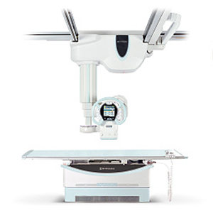
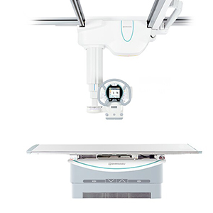
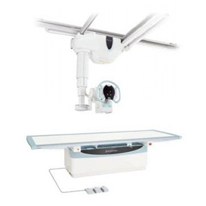
RADspeed Pro EDGE Package
Maximum Clinical Utilization
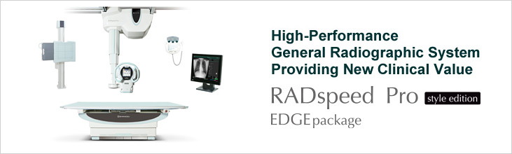
The RADspeed style edition Pro EDGE package is
the top-of the line General
Radiography System in Shimadzu RADspeed family. The Pro EDGE features a variety of the latest
cutting-edge applications like Tomosynthesis, Speed Stitch or Dual Energy Subtraction.
Providing New Clinical Value
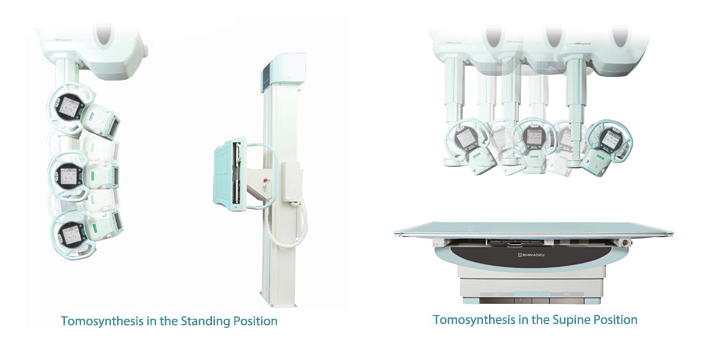
Tomosynthesis (Digital Multislice Tomography)
(*)
Tomosynthesis is a new digital imaging technology that combines cone-beam CT reconstruction with
digital image processing. It allows images of any cross section to be obtained easily from volume data
acquired from a single tomographic scan.
- Flexible Examinations with Freedom in Choosing Body Positions
This allows
images to be obtained with loads applied in the standing position, or in the supine position on
a table. Consequently, it can be used to obtain images of the elbow or knee in the bent
position, which is difficult using CT.
- Display of Oblique Cross Sections
Tilting the tomosynthesis cross section
slightly from horizontal improves the visibility of spines, hip joints, and other areas that are
not parallel to the tabletop.
T-smart (**)
T-smart”,
Tomosynthesis-Shimadzu Metal Artifact Reduction Technology,
is our latest and highest grade tomosynthesis technology evolved further to reduce the metal artifacts
with iterative reconstruction method. T-smart is useful especially for the prestigious orthopedic
customers who are treating a lot of joint replacements with metal implants because T-smart is effective
to see the adhesion degree between bone and implant.
Speed Stitch (Auto stitching of
long
view
images)
The X-ray tube swings and the FPD moves automatically to capture image data.
The captured image data is then automatically stitched together in the DR system. This makes it easy
to create long images that extend across larger areas of the body in the anteroposterior direction.
Speed Stitch function is available both in supine position and in standing position.
Dual Energy Subtraction (**)
By taking successive high and low voltage images
and applying a calculation process, soft-tissue images and bone images can be viewed separately.
Shadows of nodes obscured by ribs can be rendered in soft-tissue images, or calcification can be
rendered in bone images.
Sophisticated Functionality Makes It Even Easier to Operate
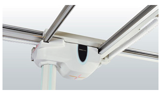
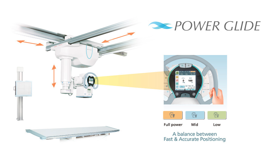
New Power Assist Function for Superb Operability and Throughput
The newly-developed POWER GLIDETM will assist your X-ray tube positioning by motors and makes it
quick
and extremely light, just like “Gliding in Air”. It will reduce technologists’ burdens and increase
patient
throughput.
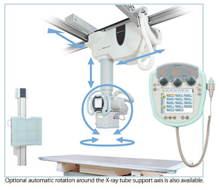 Revolutionary Auto-Positioning Feature Allows the Operator to
Focus On Patient Care
Revolutionary Auto-Positioning Feature Allows the Operator to
Focus On Patient Care
The auto-positioning feature is interlocked with the APRs.
This function moves the ceiling-mounted X-ray tube support to any desired position at the press of a
single button and can automatically set the X-ray tube angle. Effortless tube positioning allows the
operator to focus on patient care. Naturally, manual operation is also possible to make fine
positioning corrections extremely simple.
Pressing a single button on the remote control smoothly moves the ceilingmounted X-ray tube
support to preregistered positions. Movement stops immediately after the remote control button is
released. Up to two remote control units can be used.
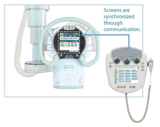
APRs Synchronized with the X-Ray High Voltage Generator
Radiography parameters can be changed beside the patient as well as on the wall-mounted
console in the control room. The operator can prepare for radiography without leaving the patient.
This sophisticated synchronization of the X-ray tube support and X-ray high voltage generator
effectively exploits the convenience of dual consoles.
New Ways to Reduce Patient Exposure
 Auto-Filtering Feature Automatically Switches to the Optimal
Filter for Each Selected Protocol (*)
Auto-Filtering Feature Automatically Switches to the Optimal
Filter for Each Selected Protocol (*)
Select a protocol to suit the type of
examination, and the filter in the collimator will change in accordance with the protocol. This
ensures the correct filter is always automatically selected.
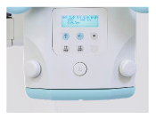
Removable Grid
Remove the grid during pediatric radiography to reduce patient
exposure. The type of grid inserted is displayed on the integrated console and on the LCD on the
ceiling-mounted X-ray tube support.
Dose-Area Product Meter / Calculated Dose (*)
For dose monitoring, either a
Dose-Area Product Meter (DAP) or a Calculated Dose is available. The DAP measures the actual dose
and displays it on the DR operator’s console. The Calculated Dose displays the expected dose, in
advance of the exposure, based on the radiography parameters and the distance to the patient. After
the exposure, the calculated dose, based on the actual exposure arameters, is displayed. With either
option, the resulting exposure parameters and dose are displayed and can be sent to the RIS/PACS
system.
Orderly Cable Management (*)
Shimadzu provides a tractable cable management system along
the
ceiling
rails that supports smooth positioning.
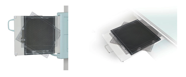
FPD Rotation Tray (*)
The FPD tray can be rotated 90 degree to change the
orientation of 14 by 17 FPD.
(*) Option
(**) Option for DR-ID911SE only


 ©
- CMS Imaging, Inc. All Rights
Reserved
©
- CMS Imaging, Inc. All Rights
Reserved