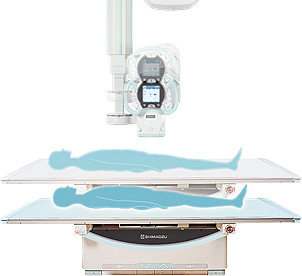
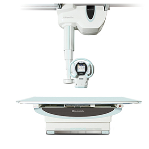
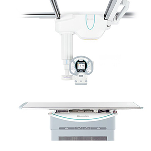
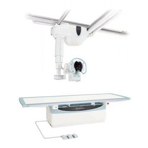
The focal point of the X-ray tube unit moves up and down matching the vertical positioning of the
X-ray Bucky stand and Bucky table. This allows the operator to attend the patient in a standing
position while positioning the equipment. For a table study, the X-ray tube automatically moves to a
preset SID, enabling accurate and fast positioning.
Vertical tracking with Bucky stand
Vertical tracking with Bucky table
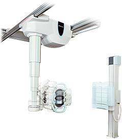
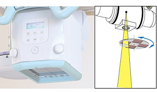
The Auto-Positioning feature is synchronized with the anatomical program. This function moves the
ceiling-mounted X-ray tube support to any desired position at the press of a single button and can
automatically set the X-ray tube angle. This effortless tube positioning allows the operator to
focus on patient care.
RADspeed Pro seamless Auto-Positioning movement
Superb Functionality Meet Various Clinical Needs
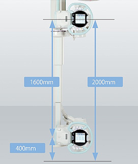
X-ray tube support vertical range of 63 inches (1,600 mm) ensures sufficient SID when examining
supine patients and low focal point radiography of standing patients. This support also rotates on
the vertical and horizontal axis in addition to fixed positioning at any desired angle, enabling
fast positioning at complex angles for orthopedic applications.
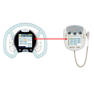
Radiography parameters and
techniques can be changed beside the patient as well as on the wall-mounted console in the control
room. The operator can prepare any radiographic exams without leaving the patient.
Sufficient vertical travel
Synchronized screen between X-ray
tube support and the console
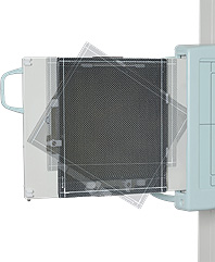
Customize Your Radiographic Room
The RADspeed Pro is customizable radiographic system depending on the customer’s needs. Easy
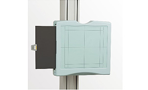
operation FPD rotation tray maximize the use of 14x17inches detector and it is a cost-effective
solution in the X-ray department. The tractable cable management system organizes the X-ray tube
support cables and provides your radiographic room with clean appearance.
FPD rotation tray for 14x17inches detect
Cable tracker for clean appearance
Comprehensive Dose Reduction and Management
The RADspeed Pro built-in dose management features help dose reduction efficiently. The collimator’s
auto-filtering feature automatically switches to an optimal beam hardening filter based on the
selected APR and reduce low energy X-ray spectrum which is mostly unnecessary for general
radiographic imaging. The built-in calculated dose display function shows the expected dose, in
advance of the exposure, based on the X-ray parameters and the distance to the patient. The
calculated dose value can be sent to the DR console and the RIS/PACS system as dose recording
purpose. (DAP meter is available as an option.) Removing the grid during radiography allows reducing
the exposure dose level in pediatric and orthopedic applications.
Automatic filter selection based on selected APR
Removable grid for pediatric exams
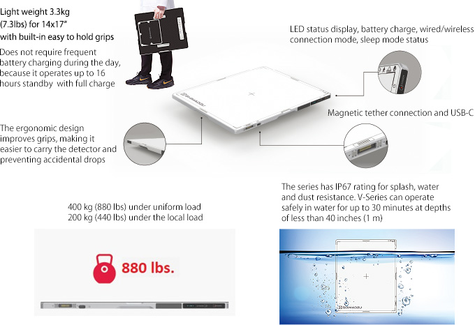
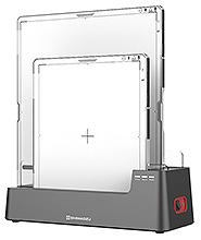
Experience New Clinical Values with an Innovative DR System
The elegance of the new design combines the robustness of product while creating a sophisticated,
powerful and practical detector in any hospital environment. V series detector is suitable for use
on the stand and table tray, table-top or cross-table exams, and ER or surgery room environment.
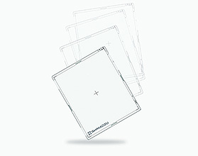
Drop resistance 1m (40 inch) height
Convenient charging Cradle for safe storing and easy charging
V1417 (14 x 17 inches) Versatile use 6.9lbs. (3.15kg)
V1717 (17 x 17 inches) Chest stand, Trauma, and Surgery 8.0lbs.
(3.65kg)
V1012 (10 x 12 inches)) Extremities and Orthopedics 4.3lbs.
(1.95kg)
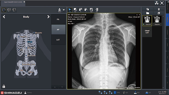
Designed with the user in mind, the digital control console offers easy navigation and touch
screen features to simplify procedures and improve workflow.
Automatically Linked
Radiography X-Ray Exposure Field (*Option) The collimator X-ray exposure field
is automatically linked to the exposure area size selected in the DR
system.
Verify the Patient Name in the Examination Room (*Option) The
patient name and ID number registered in the DR system are displayed on the X-ray tube support,
which makes it easy to verify patient information


 ©
- CMS Imaging, Inc. All Rights
Reserved
©
- CMS Imaging, Inc. All Rights
Reserved